


| Issue |
J Oral Med Oral Surg
Volume 32, Number 2, 2026
|
|
|---|---|---|
| Article Number | 11 | |
| Number of page(s) | 6 | |
| DOI | https://doi.org/10.1051/mbcb/2026014 | |
| Published online | 04 June 2026 | |
Case Report
A wolf in sheep’s clothing: malignant transformation of Ameloblastic Fibroma
1
Department of Oral and Maxillofacial Clinical Sciences, Faculty of Dentistry, Universiti Malaya, 50603 Kuala Lumpur, Malaysia
2
Stomatology Unit, Institute of Medical Research, National Institute of Health, Setia Alam, 40170 Shah Alam, Selangor, Malaysia
3
Department of Oral and Maxillofacial Surgery, National Cancer Institute, Precinct 7, 62250 Putrajaya, Malaysia
* Correspondence: This email address is being protected from spambots. You need JavaScript enabled to view it.
Received:
6
October
2025
Accepted:
26
March
2026
Abstract
With just over 100 cases reported to date, ameloblastic fibrosarcoma (AFS) is a rare and aggressive malignant odontogenic tumour that can arise de novo or develop from the malignant transformation of Ameloblastic Fibroma (AF). We report the case of a 28-year-old Malay lady who presented with a progressively enlarging, painless swelling over the left mandible, causing facial asymmetry and masticatory difficulties. Radiographic investigation revealed an expansile lesion with thin remaining cortical bone at the lower border of mandible. An incisional biopsy suggested AF, leading to surgical enucleation with peripheral ostectomy and Carnoy’s solution application. Postoperative histopathological examination, however, unexpectedly revealed areas of AFS within the lesion. Following a multidisciplinary consultation, close surveillance was recommended in view of clear surgical margins with no residual disease. The patient has completed 6 months of follow-up to date with no signs of recurrence. This case illustrates the difficulty of distinguishing AF from early-stage AFS and the need for long-term monitoring due to its malignancy potential. While radical surgery is typically the go-to treatment for AFS, this case suggests a more conservative approach with close monitoring may sometimes be appropriate. Nevertheless, further research is needed to develop clear guidelines for managing this rare malignancy.
Key words: Odontogenic tumour / Ameloblastic Fibroma / Ameloblastic Fibrosarcoma / malignant transformation / multidisciplinary
© The authors, 2026
 This is an Open Access article distributed under the terms of the Creative Commons Attribution License (https://creativecommons.org/licenses/by/4.0), which permits unrestricted use, distribution, and reproduction in any medium, provided the original work is properly cited.
Introduction
Odontogenic tumours are a diverse group of neoplastic growths that arise from the tissues involved in tooth development and the periodontal apparatus of the jaw. In the latest World Health Organization (WHO) classification, odontogenic tumours are classified into two main categories, benign and malignant tumours. Benign odontogenic tumours are further subdivided based on their histogenetic origin into epithelial, mesenchymal or mixed type [1]. Ameloblastic Fibroma (AF) falls under the category of benign mixed epithelial and mesenchymal origin, whereas ameloblastic fibrosarcoma (AFS) is its malignant counterpart [1–5]. AFS is characterized by a benign epithelial component alongside a mesenchymal component that has undergone malignant changes [6]. Approximately 50% of AFS cases arise de novo, while the remaining cases result from malignant transformation of AF [7]. A 2018 systematic review discovered that AF can undergo malignant transformation years after the initial diagnosis with the mean transformation time from the initial diagnosis of AF to development of AFS stands at 55.1 months [8]. Unfortunately, very few articles have documented the transformation of AF into AFS within the same patient, with just over 100 cases of AFS reported in the literature [5]. Therefore, this rare case report aims to contribute to the existing literature and underscore the need for further research. Decision making algorithm is shown in Figure 1.
Case report
A healthy 28-year-old Malay woman with no significant past medical, surgical, or family history presented to our department with a slow-growing swelling of the left side of lower jaw of eight months' duration. She denied any history of tobacco or alcohol use. The lesion, initially described as the size of a peanut, gradually increased in size, resulting in significant facial asymmetry and chewing difficulty. Our initial differential diagnoses included ameloblastoma, odontogenic myxoma, dentigerous cyst, odontogenic keratocyst, central odontogenic fibroma, and a possible low-grade sarcoma. Extraoral examination revealed a large, diffuse swelling measuring 6 cm × 7 cm in size, extending from the left mandibular body to the preauricular region, with normal overlying skin (Fig. 2A). No palpable cervical lymphadenopathy or paraesthesia was noted. Intraorally, there was a large exophytic growth at the posterior mandible measuring around 5 cm × 6 cm in size with significant buccolingual expansion. The mass interfered with occlusion and was associated with surface ulceration and bite marks (Fig. 2B). An incisional biopsy was performed and the histopathology examination (HPE) report was suggestive of AF. Further investigations with orthopantomogram (OPG) and computed tomography (CT) were carried out. OPG demonstrated a well-defined radiolucent lesion in the left posterior mandible, extending from the first molar to the ramus. Significant features included thinning of the inferior cortical border and association with an impacted mandibular molar. The inferior alveolar canal was displaced inferiorly but remained intact (Fig. 2C). CT revealed a large expansile lesion measuring approximately 5.6 × 6.7 × 7.0 cm, involving the mandibular body, angle, and ramus (Figs. 2D and 2E). While there was significant cortical thinning with buccolingual expansion, the CT confirmed the absence of cortical perforation, adjacent soft-tissue invasion, increased vascularity, or cervical lymphadenopathy.
The patient was then subjected to enucleation of the lesion at the left mandible using both intraoral and extraoral approaches. Following enucleation, a peripheral ostectomy and application of Carnoy’s solution was performed. The defect was packed with a ribbon gauze soaked in white head’s varnish and a reconstruction plate was placed to provide stability to the thin residual lower border of the mandible (Figs. 2F and 2G).
Despite the lesion’s large size, primary closure was successfully achieved with the remaining soft tissue. Her post-operative recovery was uneventful, and she was discharged home on post-operative day 6.
Unexpectedly, her post-operative HPE revealed AF with focal areas of AFS. The HPE displayed multilobulated odontogenic neoplasm composed of fibromyxoid connective tissue resembling dental papilla intermixed with scattered odontogenic epithelium. The mesenchymal component contained ovoid, spindle to stellate-shaped cells with vesicular to hyperchromatic areas whereas the epithelial component was composed of bilaminar cuboidal to low columnar odontogenic epithelial cells arranged in small nests, narrow cords and strands with hyperchromatic palisaded nuclei with reversed polarity. In certain areas, malignant transformation of the mesenchymal cells was observed, characterised by hypercellularity, moderate degree of cellular and nuclear pleomorphism, and increased mitosis. The areas occupy around 35% of the tumour area. Ki-67 proliferative index was also higher at the hypercellular areas, around 30–40% (Figs. 3F and 3G).
Her case was then discussed at the multidisciplinary clinic (MDT). The consensus was for close surveillance with monthly review for the first 2 yr followed by lifelong periodic reviews. The recommended investigations included a baseline postoperative CT scan of the thorax, abdomen, and pelvis (TAP), magnetic resonance imaging (MRI) of the head and neck at three months post operative and periodic OPG imaging at three-month intervals. Imaging demonstrated no evidence of distant metastasis or residual tumour (Figs. 3C, 3H, 3I and 3J). The patient remains disease free at 6 months of follow-up with no signs of recurrence and has resumed normal daily activities (Figs. 3A, 3B, 3D and 3E). The timeline of events are summarised in Table I.
Discussion
AFS is a rare and locally aggressive malignant tumour which accounts for less than 5% of all odontogenic tumours [9]. It was first described by Heath in 1887 [6]. AFS primarily affects young adults, with a mean reported age of 30 yr and has a male to female ratio of 1.6:1, consistent with the age of our 28-year-old patient. The posterior mandible is the most frequently affected site [5]. Clinically, AFS presents as a swelling that can be painful or painless, occasionally accompanied by paresthesia or dysesthesia. Radiologically, it appears as a radiolucent mass with ill-defined borders and cortical bone perforation. Histologically, it can be difficult to differentiate AFS from its benign counterpart AF as the mesenchymal component AFS displays stepwise progression towards malignancy. This progression is characterized by marked tumour cellularity, nuclear polymorphism, hyperchromatism and increased mitotic figures [7]. Because of its aggressive nature, radical surgical excision with clear margins is the preferred treatment for AFS. Cervical lymph node dissection is not required because sarcoma spreads via vascular channels with no lymphatic metastasis [6]. The use of adjuvant radiotherapy and chemotherapy on the other hand has often been disputed with varying preferences among surgeons and oncologists. Delivering high dose radiation therapy of 50–60 Grays (Gy) directly to the tumour has the capability to decrease recurrence and prevent metastasis. Similarly, adjuvant chemotherapy using cyclophosphamide and fluorouracil has also demonstrated some success, but no standardized treatment protocols have yet to be established [6,9,10].
Our decision to manage this case conservatively was based on the initial HPE diagnosis of AF as well as the clinico-radiographical features of the lesion. Evidences have recommended conservative surgery as the first-line treatment for AF, particularly in young patients [6,8,11,12]. While we acknowledge that incisional biopsies carry a risk of sampling error, potentially missing focal sarcomatous elements of a developing AFS, radical resection of a histologically and clinico-radiographically benign lesion should be reconsidered to prevent unnecessary surgical morbidity especially cosmetic defects in young patients.
The clinical presentation in our patient was consistent with that of AF, typically manifesting as a painless jaw swelling. Many AF cases are asymptomatic and are often detected incidentally on radiographic examination The radiographic features of AF typically include a well-defined unilocular or multilocular radiolucency with sclerotic borders and may also be associated with unerupted or displaced teeth [3], as observed in the present case. Due to its indolent nature, AF does not infiltrate bone and is therefore easily separable intraoperatively, as demonstrated in the present case, where the tumour remained well encapsulated and was completely excised. Hence, it is treated more conservatively compared to ameloblastoma, with total enucleation and curettage the standard treatment approach. Nevertheless, it does still have a recurrence rate of up to 20% with potential risk of malignant transformation. Therefore, these patients require regular long-term follow-up [2].
We performed peripheral ostectomy and applied Carnoy’s solution to minimize residual microscopic tumour and prevent recurrence. The adjunctive use of Carnoy’s, which is a chemical cauterizing and fixative agent, following conservative excision has been shown to significantly reduce recurrence rates in benign but aggressive odontogenic tumours such as ameloblastoma and odontogenic cysts. A published systematic review showed enucleation with Carnoy’s had a recurrence rate of only 16%, compared to 30% for enucleation alone [13].
Given the high local recurrence rate of AFS which is reported between 23.9% and 37%, a close follow-up protocol is essential [6,11,12,14]. We opted for a monthly clinical review for the first 2 yr because evidence suggests that the majority of AFS recurrences occur within this critical 12-to-24-month postoperative window [6,8,10–12]. This frequent schedule is particularly indicated in cases of secondary AFS, as suspected in our patient, where the malignancy arises from a pre-existing AF carry a significantly higher recurrence risk of 50% compared to de novo lesions [4]. We performed MRI three months post operatively on the recommendation of our head and neck radiology team, as it provides superior soft-tissue detail necessary to differentiate tumor recurrence from expected postoperative changes. Additionally, while distant metastasis in AFS is relatively rare (occurring in 4.5% to 9% of cases) [5,8,15–17], a CT TAP (Thorax, Abdomen, and Pelvis) is necessary for baseline staging and systemic surveillance. This is justified by the fact that the lungs are the most common site of distant spread (accounting for 66.7% of metastases) [15] and the overall mortality rate of the disease remains significant at approximately 19% to 25% [8,15,18]. OPG will be performed 3 monthly as it is a low-cost imaging modality that enables the monitoring of mandibular bone integrity and plate fixation while facilitating the early detection of radiolucent changes indicative of local recurrence with minimal radiation exposure.
In summary, the diagnostic discrepancy between an initial incisional biopsy reporting AF and a final post-operative diagnosis of AFS could be attributed to sampling error, the focal nature of malignant transformation within the mesenchymal component or an early malignant changes in a lesion initially diagnosed as AF [10,15,19]. Because sarcomatous changes are often not distributed uniformly throughout the tumour volume, limited biopsy specimens may fail to capture malignant areas, a challenge documented in reports by Mothlagh et al., Munisekhar et al., and Lai et al. [11,16,19]. To prevent such misdiagnosis, clinicians must ensure thorough sampling from multiple representative areas and maintain high suspicion when clinico-radiographic features such as ill-defined margins or cortical bone perforation indicate an aggressive tumour not reflected in the initial biopsy [15,19]. Furthermore, the routine application of immunohistochemical markers like Ki-67 and p53 should be considered, as AFS typically exhibits significantly higher proliferative indices in its mesenchymal component compared to AF [15,19]. Given the documented potential for AF to transform into AFS following conservative treatment, as reported in cases by Kobayashi et al., Hu et al., and Ramakrishnan et al. [6,10,20], more aggressive initial surgical resection may be warranted to mitigate the risks of missed malignancy or future sarcomatous recurrence.
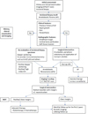 |
Fig. 1 Decision making algorithm. |
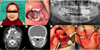 |
Fig. 2 Pre-operative assessment and surgical management of the patient. (A) Pre-operative extraoral appearance; (B) Pre-operative intraoral appearance; (C) OPG showing a radiolucent lesion at the left mandible with an impacted molar; (D) – (E) Axial and coronal CT scan slices demonstrating a large expansile bony lesion at the left mandible with cortical thinning; (F) Intraoral approach exposing the tumour and associated impacted tooth; (G) Reconstruction plate placed at the lower left border of the mandible following enucleation of the tumour. |
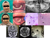 |
Fig. 3 Post-operative follow-up and histopathological findings. (A) Extraoral appearance of the patient at post-operative 1 month; (B) Intraoral condition of the surgical site at post-operative 1 month; (C) Post-operative OPG displaying an intact reconstruction plate with screws; (D) Extraoral appearance of the patient at post-operative 6 months; (E) Intraoral condition of the surgical site at post-operative 6 months; (F) Haematoxylin & Eosin, 20x magnification: Photomicrograph shows cords, islands and nests of benign odontogenic epithelial cells (white arrowhead) in the background of hypercellular stroma resembling dental papilla (black arrowhead). These features are consistent with Ameloblastic Fibroma; (G) Haematoxylin & Eosin, 40x magnification: Photomicrograph shows malignant ectomesenchyme displaying hypercellularity with cells having moderate cellular and nuclear pleomorphism and increased mitotic activity (black arrowhead). The epithelial component appears cytologically bland (white arrowhead); (H), (I) and (J) Respective coronal, axial and sagittal views of the MRI taken 3 months postoperatively showing no signs of recurrence or residual tumour. |
Chronological timeline of events.
Funding
No funding was obtained for this case report.
Conflicts of interest
There is no conflict of interest to declare.
Data availability statement
All the data relevant to the publication are presented within the article.
Ethics approval
No approval was required for this case report.
Informed consent
Patient’s consent was obtained.
References
- Soluk-Tekkesin M, Wright JM. The World Health Organization classification of odontogenic lesions: a summary of the changes of the 2022 (5th) edition. Turk Patoloji Derg 2022:38: 168–184. [Google Scholar]
- Binthiyaz T, Devados P. Ameloblastic Fibroma of mandible- a case report. Int J Oral Health Dent;2024:10;34–36. [Google Scholar]
- McDonald JS. Tumors of the oral soft tissues and cysts and tumors of bone. In: McDonald and Avery's Dentistry for the Child and Adolescent, 10th edn. St. Louis, MO: Mosby. 2016;603–626. [Google Scholar]
- Nasir A, Khare A, Ali I, Khan MI. Ameloblastic Fibroma: a case report. J Oral Maxillofac Pathol 2023:27;S60–S63. [Google Scholar]
- Barakeh D, Alsolami A, Abedalthagafi M. Ameloblastic Fibrosarcoma of the jaw: case report, genetic profiling, and literature review. Case Rep Oncol 2023:16;1293–1299. [Google Scholar]
- Ramakrishnan DS, Gouthaman SS, Muthusekhar MR. Ameloblastic Fibrosarcoma transformation from Ameloblastic Fibroma. Natl J Maxillofac Surg 2022 13;S145–S149. [Google Scholar]
- Bertoni F, Del Corso G, Bacchini P, Marchetti C, Tarsitano A. Ameloblastic Fibrosarcoma of the mandible evolving from a prior Ameloblastic Fibroma after two years: an unusual finding. Int J Surg Pathol 2016:24;656–659. [Google Scholar]
- Chrcanovic BR, Brennan PA, Rahimi S, Gomez RS. Ameloblastic Fibroma and ameloblastic fibrosarcoma: a systematic review. J Oral Pathol Med 2018:47;315–325. [Google Scholar]
- Liu S, Li H, Dong Y, Zhang D. Ameloblastic Fibrosarcoma of the maxilla arising in an old woman, a rare case report and literature review. BMC Oral Health 2024;24:743. [Google Scholar]
- Hu YY, Deng MH, Yuan LL, Niu YM. Ameloblastic Fibrosarcoma of the mandible: a case report and mini review. Exp Ther Med 2014;8;1463–1466. [Google Scholar]
- Munisekhar MS, Shylaja S, Kumar RV, Rao KA, Patil SR, Alam MK. Ameloblastic Fibrosarcoma-a rarity? JPRAS Open 2019;21;56–62. [Google Scholar]
- Hascoet E, Le Cléac’h S, Taugeron F, Bertin H, Longis J, Piot B, et al. Ameloblastic Fibrosarcoma of the mandible: case report and literature review. Med Buccale Chir Buccale 2017:23;164–168. [Google Scholar]
- Lau SL, Samman N. Recurrence related to treatment modalities of unicystic ameloblastoma: a systematic review. Int J Oral Maxillofac Surg 2006;35;681–690. [Google Scholar]
- Soltany A, Asaad G, Daher R, Dayoub M, Khalil A, Alshehabi Z. A maxillary Ameloblastic Fibrosarcoma tumor: a rare case report from Syria. J Surg Case Rep 2020 2020;rjaa255. [Google Scholar]
- Servato JPS, de Faria PR, Ribeiro CV, Cardoso SV, de Faria PR, Dias FL, et al. Ameloblastic Fibrosarcoma: a case report and literature review. Braz Dent J 2017:28;262–272. [Google Scholar]
- Lai J, Blanas N, Higgins K, Klieb H. Ameloblastic Fibrosarcoma: report of a case, study of immunophenotype, and comprehensive review of the literature. J Oral Maxillofac Surg 2012:70;2007–2012. [Google Scholar]
- Loya-Solis A, González-Colunga KJ, Pérez-Rodríguez CM, Ramírez-Ochoa NS, Ceceñas-Falcón L, Barboza-Quintana O. Ameloblastic Fibrosarcoma of the mandible: a case report and brief review of the literature. Case Rep Pathol. 2015 2015;245026. [Google Scholar]
- Gilani SM, Raza A, Al-Khafaji BM. Ameloblastic Fibrosarcoma: a rare malignant odontogenic tumor. Eur Ann Otorhinolaryngol Head Neck Dis 2014:131;53–56. [Google Scholar]
- Motlagh MF, Mostafazadhe S, Motlagh FF. Low-grade Ameloblastic Fibrosarcoma of the mandible: a case report with initial misdiagnosis. Int J Clin Oral Maxillofac Surg 2025:11;58–62. [Google Scholar]
- Kobayashi K, Murakami R, Fujii T, Hirano A. Malignant transformation of Ameloblastic Fibroma to ameloblastic fibrosarcoma: case report and review of the literature. J Cranio-Maxillofac Surg 2005:33;352–355. [Google Scholar]
Cite this article as: Singh N, Lim D, Goh Pei En J, Yusoff MN, Thangavalu L. 2026. A wolf in sheep’s clothing: malignant transformation of Ameloblastic Fibroma. J Oral Med Oral Surg. 32: 11. https://doi.org/10.1051/mbcb/2026014
All Tables
All Figures
|
Fig. 1 Decision making algorithm. |
| In the text | |
|
Fig. 2 Pre-operative assessment and surgical management of the patient. (A) Pre-operative extraoral appearance; (B) Pre-operative intraoral appearance; (C) OPG showing a radiolucent lesion at the left mandible with an impacted molar; (D) – (E) Axial and coronal CT scan slices demonstrating a large expansile bony lesion at the left mandible with cortical thinning; (F) Intraoral approach exposing the tumour and associated impacted tooth; (G) Reconstruction plate placed at the lower left border of the mandible following enucleation of the tumour. |
| In the text | |
|
Fig. 3 Post-operative follow-up and histopathological findings. (A) Extraoral appearance of the patient at post-operative 1 month; (B) Intraoral condition of the surgical site at post-operative 1 month; (C) Post-operative OPG displaying an intact reconstruction plate with screws; (D) Extraoral appearance of the patient at post-operative 6 months; (E) Intraoral condition of the surgical site at post-operative 6 months; (F) Haematoxylin & Eosin, 20x magnification: Photomicrograph shows cords, islands and nests of benign odontogenic epithelial cells (white arrowhead) in the background of hypercellular stroma resembling dental papilla (black arrowhead). These features are consistent with Ameloblastic Fibroma; (G) Haematoxylin & Eosin, 40x magnification: Photomicrograph shows malignant ectomesenchyme displaying hypercellularity with cells having moderate cellular and nuclear pleomorphism and increased mitotic activity (black arrowhead). The epithelial component appears cytologically bland (white arrowhead); (H), (I) and (J) Respective coronal, axial and sagittal views of the MRI taken 3 months postoperatively showing no signs of recurrence or residual tumour. |
| In the text | |
Current usage metrics show cumulative count of Article Views (full-text article views including HTML views, PDF and ePub downloads, according to the available data) and Abstracts Views on Vision4Press platform.
Data correspond to usage on the plateform after 2015. The current usage metrics is available 48-96 hours after online publication and is updated daily on week days.
Initial download of the metrics may take a while.