


Fig. 3
Download original image
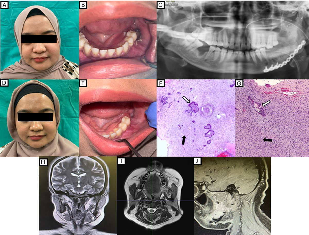
Post-operative follow-up and histopathological findings. (A) Extraoral appearance of the patient at post-operative 1 month; (B) Intraoral condition of the surgical site at post-operative 1 month; (C) Post-operative OPG displaying an intact reconstruction plate with screws; (D) Extraoral appearance of the patient at post-operative 6 months; (E) Intraoral condition of the surgical site at post-operative 6 months; (F) Haematoxylin & Eosin, 20x magnification: Photomicrograph shows cords, islands and nests of benign odontogenic epithelial cells (white arrowhead) in the background of hypercellular stroma resembling dental papilla (black arrowhead). These features are consistent with Ameloblastic Fibroma; (G) Haematoxylin & Eosin, 40x magnification: Photomicrograph shows malignant ectomesenchyme displaying hypercellularity with cells having moderate cellular and nuclear pleomorphism and increased mitotic activity (black arrowhead). The epithelial component appears cytologically bland (white arrowhead); (H), (I) and (J) Respective coronal, axial and sagittal views of the MRI taken 3 months postoperatively showing no signs of recurrence or residual tumour.
Current usage metrics show cumulative count of Article Views (full-text article views including HTML views, PDF and ePub downloads, according to the available data) and Abstracts Views on Vision4Press platform.
Data correspond to usage on the plateform after 2015. The current usage metrics is available 48-96 hours after online publication and is updated daily on week days.
Initial download of the metrics may take a while.