| Issue |
J Oral Med Oral Surg
Volume 32, Number 1, 2026
|
|
|---|---|---|
| Article Number | 9 | |
| Number of page(s) | 9 | |
| DOI | https://doi.org/10.1051/mbcb/2026009 | |
| Published online | 13 May 2026 | |
Systematic Review
Immunoexpression of P53 in ameloblastomas: a systematic review
Department of Oral Surgery, International Faculty of Dental Medicine, International University of Rabat, Morocco
* Correspondence: This email address is being protected from spambots. You need JavaScript enabled to view it.
Received:
28
January
2025
Accepted:
27
March
2026
Abstract
Objective: The objective of this systematic review is to evaluate the available evidence on P53 immunoexpression in ameloblastomas and to investigate its potential association with tumor behavior in order to clarify its diagnostic significance and prognostic implications. Methods: Following PRISMA guidelines, research in PubMed, Web of Science, Scopus, and ScienceDirect databases yielded 17 studies meeting the inclusion criteria.
Results: Results indicate that P53 overexpression is frequent in ameloblastomas, particularly in solid and multicystic types, and correlates with increased tumor aggressiveness and recurrence. A relationship was observed between P53 expression and other key molecular markers, including Bcl-2 and Ki-67, emphasizing its role in disrupted apoptotic pathways. Conclusions: These findings support the potential of P53 as a prognostic marker and suggest further research into targeted therapies for better management of ameloblastomas.
Key words: P53 / ameloblastoma / systematic review
© The authors, 2026
 This is an Open Access article distributed under the terms of the Creative Commons Attribution License (https://creativecommons.org/licenses/by/4.0), which permits unrestricted use, distribution, and reproduction in any medium, provided the original work is properly cited.
Introduction
Ameloblastoma (AMB) is a benign odontogenic tumor that accounts for approximately 10% of all craniofacial tumors. Although histologically benign, it is characterized by persistent growth, local invasiveness, and a high risk of recurrence, with recurrence rates reaching up to 70%, and in some cases, they may transform into malignant forms [1,2]. The tumor’s clinical behavior is influenced not only by its histological subtype but also by underlying molecular alterations [3,4].
Among the molecular pathways involved in ameloblastoma pathogenesis, the P53 tumor suppressor gene has emerged as a key focus of interest due to its critical role in cell cycle regulation, DNA repair, and apoptosis. Overexpression of P53 protein has been reported in various odontogenic tumors, including ameloblastomas, and appears to be associated with more aggressive histological subtypes. [5,6]
Several narrative reviews and original studies have addressed P53 expression in ameloblastomas, sometimes in relation to other markers such as Ki-67, Bcl-2, or MDM2 [3,6–8]. However, these studies remain limited by small sample sizes, inconsistent methodologies, and a lack of standardized synthesis. To our knowledge, no systematic review has yet applied PRISMA methodology to provide a structured analysis of P53 expression across different histological variants of ameloblastomas.
The objective of this systematic review is to evaluate the available evidence on P53 immunoexpression in ameloblastomas and to investigate its potential association with tumor behavior in order to clarify its diagnostic significance and prognostic implications.
Materials and methods
This systematic review was performed in accordance with the PRISMA 2020 guidelines (preferred reporting items for systematic reviews and meta-analyses) and is registered in the PROSPERO database under the registration number CRD42022355941.
Focused question
We intended to answer the following focused question: Is there an overexpression of P53 in patients with ameloblastoma?
According to the study hypothesis, the PECOS criteria were defined as follows:
Population:
Healthy individuals without systemic diseases.
Exposure:
Patients diagnosed with ameloblastomas.
Comparison:
Ameloblastomas (AME) of varied histological typology,
Dentigerous cyst (DC),
Odontogenic keratocyst (OKC),
Adenomatoid odontogenic tumors (AOT),
Ameloblastic carcinoma (AMECA).
Outcomes:
Expression of p53 protein,
Setting:
Immunohistochemical (IHC) studies,
Polymerase chain reaction (PCR).
Study selection criteria
Inclusion criteria
Studies were considered eligible for inclusion if they met all of the following criteria:
Original research articles focusing on the expression of p53 in odontogenic tumors, particularly ameloblastomas.
Studies conducted on human tissue samples, with data obtained through IHC or PCR.
Case reports and case series providing original IHC or PCR data on p53 expression.
Articles published in English.
Studies published between 2010 and 2024.
Studies involving patients without systemic diseases or comorbidities that could influence gene expression.
Exclusion criteria
Studies were excluded if they met one or more of the following criteria:
Articles focusing primarily on the treatment (surgical, radiotherapeutic, or medical) of ameloblastomas, without p53 expression data.
Studies conducted on animal models or in vitro cell cultures.
Review articles, letters to the editor, or conference abstracts without original data.
Descriptive case reports without molecular analysis.
Articles published in languages other than English.
Studies involving patients with systemic conditions (e.g., autoimmune diseases, cancer syndromes) potentially affecting molecular markers.
Studies not reporting quantitative or qualitative data on p53 expression using IHC or PCR methods.
Search strategy
The initial search was performed online by two independent reviewers (Y.A. and S.A.) from the start of the study up to November 2024. Duplicate records were then removed. Preliminary research was conducted in databases including PubMed, Scopus, Web of Science, ScienceDirect, and the Cochrane Library to identify relevant systematic reviews and articles. These searches helped establish terms and synonyms related to the primary concepts of interest (P53 and amelobastoma) and to evaluate and develop the most optimal search strategy. In addition to database searches, manual search was also conducted.
The following strategy was used: (ameloblastoma or ameloblastomas) and (p53 antigen or P53 tumor suppressor protein or cellular tumor antigen p53 or oncoprotein p53).
Study selection
The initial selection of records from the first hit was conducted by independently reviewing their titles. In the second round, abstracts of the remaining records were reviewed, followed by full-text assessments in the third round. All records underwent independent evaluations by the same reviewers to determine their eligibility. Any disagreements between the two reviewers during the abstract and full-text screening were resolved through consultation with a third reviewer (S.C.). Studies that did not satisfy the inclusion criteria were excluded from the analysis.
Data collection process
For each study included in the final review, relevant data were independently extracted by three reviewers (Y.A., S.A., N.A.). Any discrepancies among the three reviewers during the data extraction process were resolved through discussion with a fourth reviewer (S.C.). The extracted information included the first author’s name and the year of publication, methods used (immunohistochemical technique or PCR technique), types of lesions, number of samples, and reported outcomes. All extracted data were organized in tables using Office software by two reviewers (Y.A., G.E.). The data extraction forms, along with the findings from each included study, were collectively reviewed to ensure the accuracy and reliability of the process.
Quality assessment of the included articles
A risk of bias assessment for the included studies was performed in accordance with the guidelines outlined in the Cochrane Handbook for Systematic Reviews of Interventions (2008), Chapter 8: Assessing Risk of Bias in Included Studies [9].
An adapted checklist based on the Cochrane framework was used to evaluate the methodological quality of each study. The tool comprised five key domains:
Clearly defined study objective,
Adequate description of inclusion criteria,
Clearly defined protocol,
Adequate statistical analysis,
Clearly described main outcomes.
Each domain was rated as follows:
Yes: Low risk of bias
No: High risk of bias
Uncertain: Insufficient information or ambiguity regarding potential bias
This adaptation ensured methodological consistency across the included observational and descriptive studies.
Results
Literature search
Figure 1 presents a PRISMA flow diagram illustrating the study selection process based on the specified inclusion criteria. An extensive search across the selected databases yielded 101 references for potential inclusion in this systematic review. After removing 9 duplicate records, 92 unique articles remained for screening. Following title and abstract screening, 55 articles were excluded for not meeting the inclusion criteria. The remaining 37 full-text articles were assessed for eligibility. Among these, 20 were excluded after thorough examination due to reasons such as non-human samples, lack of P53 analysis, or being review articles. Consequently, 17 studies were included in the final systematic review.
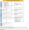 |
Fig. 1 Flow diagram. |
Main outcomes
The key findings from the included studies are summarized in Table I.
Across the 17 included studies, the majority (approximately 76%, 13 out of 17) reported moderate to high levels of P53 expression in ameloblastomas, suggesting a potential involvement in tumor pathogenesis. However, four studies reported low, absent, or statistically non-significant expression [14,18,19,21], indicating heterogeneity in the reported findings.
Regarding histological subtypes, most studies (approximately 88%, 15 out of 17) reported moderate to high levels of P53 expression in solid/multicystic ameloblastomas [7,8], whereas lower expression was more commonly observed in unicystic variants [10]. However, one study [22] presented an opposite finding, reporting higher P53 positivity in unicystic ameloblastomas compared with solid/multicystic types.
Several studies demonstrated that P53 expression was frequently higher in follicular subtypes compared with plexiform or unicystic forms [7,11].
In more aggressive variants such as basal cell ameloblastomas and ameloblastic carcinomas, very high P53 expression was reported [12,13], often accompanied by elevated levels of Ki-67 and MDM2, indicating increased proliferative potential and malignant transformation risk.
Studies that explored co-expression with apoptotic markers (Bcl-2, Bax) reported an imbalance favoring cell survival in aggressive variants [8,14], suggesting a mechanistic role for P53 in resistance to apoptosis.
Additionally, Singh et al. [15] showed comparative overexpression of P53 in odontogenic keratocysts and solid ameloblastomas, reinforcing its involvement in tumor progression. Salehinejad et al. [16] also supported the proliferative role of P53 by demonstrating its co-expression with PCNA in ameloblastomas, further linking P53 to tumor growth dynamics.
Meanwhile, Kitkumthorn et al. [6] investigated a P53 codon 72 polymorphism and its potential association with ameloblastoma susceptibility, offering additional genetic insights into tumor pathogenesis.
Moreover, Beppu et al. [17] described a rare and rapidly evolving case of ameloblastoma characterized by markedly increased P53 expression and accelerated cell proliferation, suggesting that P53 dysregulation may contribute to early tumor dedifferentiation and invasiveness in select cases.
Although not all studies reported exact quantitative percentages, the majority identified moderate to strong nuclear P53 staining in 50%–80% of tumor cells, particularly in peripheral epithelial layers.
To provide a clearer comparative overview, Table II summarizes the reported patterns of p53 expression across different histological variants of ameloblastoma, their associated molecular markers, and the potential prognostic implications described in the included studies. This synthesis facilitates the interpretation of the heterogeneity of p53 expression and its possible relevance to tumor behavior.
Features and main outcomes of the included studies.
Expression patterns of P53 and associated markers in histological subtypes of ameloblastoma.
Quality assessment
The quality assessment of the included studies was conducted based on the criteria outlined in the Cochrane Handbook for systematic reviews of interventions [9].
One study [17] was identified as having a higher risk of bias, with a “No” judgment for adequate statistical analysis and an “Uncertain” judgment for the description of main results. Three studies [15,17,18] received “Uncertain” or “No” responses regarding statistical analysis, indicating a potential moderate risk of bias in this domain.
All other studies answered “Yes” to the questions regarding a clearly defined study objective, adequate inclusion criteria, and a clearly defined protocol, reflecting a generally low risk of bias in these aspects. The question “Was an appropriate statistical analysis used?” was answered affirmatively by 13 out of the 17 studies, showing consistency in the use of robust statistical methods across most studies. Regarding the description of main results, two studies [17,18] provided unclear or “Uncertain” responses, suggesting limitations in how outcomes were reported.
Overall, among the 17 included studies, four presented potential weaknesses related to statistical analysis and reporting clarity, while the remaining 13 were judged to have a low risk of bias overall.
The risk of bias assessment for the studies is summarized in Table III.
Quality of the included studies.
Discussion
This review highlights the relevance of P53 immunoexpression as a potential prognostic marker in ameloblastomas. Majority of studies demonstrated that higher P53 levels were observed in more aggressive histological subtypes, such as Solid/Multicystic ameloblastomas (SMA) and Ameloblastic Carcinomas (AMECA). For instance, Singh et al. [15] reported the highest P53 expression in SMA, while Loyola et al. [12] found very strong nuclear staining in malignant variants, suggesting a role in tumor transformation and poor clinical behavior.
However, it is important to note that one study [22] reported higher p53 positivity in unicystic ameloblastomas compared with solid/multicystic variants, which contrasts with the general trend. Despite this, approximately 88% of the included studies (15 out of 17) demonstrated higher P53 expression in more aggressive histological subtypes such as SMA and AMECA. This discrepancy may be attributed to methodological variations among studies, including differences in antibody clones, sample sizes, and scoring criteria, which could influence staining intensity and interpretation.
Beyond its association with histological subtype, P53 appears to play a central role in the molecular pathogenesis of ameloblastomas. Co-expression with other markers such as Ki-67, Bcl-2, Bax, and MDM2 suggests that dysregulation of the P53 pathway contributes to increased cellular proliferation, apoptotic resistance, and tumor invasiveness [8,13,14]. Florescu et al. [20] similarly reported high expression of P53, Bcl-2, and Ki-67 in follicular solid ameloblastomas, reinforcing its involvement in aggressive biological behavior.
While most included studies did not provide direct outcome data such as recurrence rates or survival, indirect prognostic associations were frequently noted. High P53 expression was consistently found in variants known for their higher recurrence risk, such as follicular and solid ameloblastomas [7,12,19]. These findings highlight the potential use of P53 as a surrogate marker of aggressive clinical behavior, especially in scenarios where histological evaluation may not fully reflect the tumor’s biologic potential.
In addition, comparative studies such as that by de Vicente et al. [20] highlighted significantly stronger P53 immunoreactivity in odontogenic keratocysts and ameloblastomas versus less aggressive odontogenic lesions, underscoring the marker's relevance for tumor stratification and differential diagnosis.
From a clinical standpoint, the routine immunohistochemical evaluation of P53 expression in ameloblastomas could significantly aid in stratifying patients according to their risk profile. High levels of P53 expression, particularly in solid/multicystic or follicular variants, may indicate a greater likelihood of aggressive behavior, recurrence, or even malignant transformation. In such cases, clinicians might consider performing wider surgical excisions to ensure complete tumor removal and minimize the risk of local relapse. Furthermore, these patients may benefit from a more rigorous postoperative surveillance program, including more frequent clinical evaluations and imaging studies, to detect early signs of recurrence.
In addition, understanding the P53 expression profile could support the selection of patients for future adjuvant or targeted therapies, particularly as molecular-based treatments continue to develop. While not yet standard in clinical practice, such molecular insights could eventually complement histological assessment and improve personalized treatment planning. Thus, integrating P53 assessment into routine diagnostic workflows may enhance prognostic accuracy and help tailor management strategies for improved patient outcomes.
Nevertheless, this review also highlights current limitations in literature. Inconsistent methodologies, heterogeneous antibody clones, small sample sizes, and variable scoring systems limit the ability to generalize results. There is a clear need for larger, multicenter studies with standardized immunohistochemical protocols and outcome tracking to fully validate the prognostic significance of P53 in ameloblastomas.
Conclusion
This systematic review confirms that there is a consistent overexpression of P53 in ameloblastomas, particularly in solid/multicystic variants, compared with other odontogenic lesions. This overexpression correlates with the aggressive biological behavior and higher recurrence potential of these tumors. Therefore, P53 can be considered both a useful prognostic biomarker and a potential therapeutic target. Further standardized studies are needed to validate its clinical utility and explore targeted molecular therapies.
Acknowledgments
During the preparation of this work, the authors used ChatGPT, an AI-powered language tool, to refine grammar, correct spelling errors, and enhance sentence structure for improved clarity and readability. After utilizing this tool, the authors carefully reviewed and edited the content to ensure its accuracy and integrity. The authors take full responsibility for the scientific content and originality of this publication.
Funding
No funding was received for this study.
Conflicts of interest
The authors declare no conflicts of interest.
Data availability statement
The data supporting the findings of this study are available within the article. No additional datasets were generated or analyzed during the current study.
Author contribution statement
*Conceptualization and study design : Y Azzouz, S Chbicheb;
*Data collection and analysis : Y Azzouz, S Abidi;
*Manuscript drafting : Y Azzouz ;
*Critical revision of the manuscript : All authors.
References
- Chae MP, Smoll NR, Hunter-Smith DJ, Rozen WM. Establishing the natural history and growth rate of ameloblastoma with implications for management: systematic review and meta-analysis. PLoS One 2015;10:e0117241. [Google Scholar]
- DeVilliers P, Suggs C, Simmons D, Murrah V, Wright JT. Microgenomics of ameloblastoma. J Dent Res 2011;90:463-469. [Google Scholar]
- Effiom OA, Ogundana OM, Akinshipo AO, Akintoye SO. Ameloblastoma: Current etiopathological concepts and management. Oral Dis 2018;24:307-316. [Google Scholar]
- Hendra FN, Natsir Kalla DS, Van Cann EM, de Vet HCW, Helder MN, Forouzanfar T. Radical vs conservative treatment of intraosseous ameloblastoma: Systematic review and meta-analysis. Oral Dis 2019;25:1683-1696. [Google Scholar]
- Gimenez-Conti IB, et al. p53, Rb and cyclin D1 expression in human oral verrucous carcinomas. Cancer 1996;78:17-23. [Google Scholar]
- Kitkumthorn N, Yanatatsaneejit P, Rabalert J, Dhammawipark C, Mutirangura A. Association of p53 codon 72 polymorphism and ameloblastoma. Oral Dis 2010;16:631-635. [Google Scholar]
- Sharifi-Sistani N, Zartab H, Babakoohi S, Saghravanian N, Jamshidi S, Esmaili H, et al. Immunohistochemical comparison of the expression of p53 and MDM2 proteins in ameloblastomas and keratocystic odontogenic tumors. J Craniofac Surg 2011;22:1652-1656. [Google Scholar]
- Gadbail AR, Patil R, Chaudhary M. Co-expression of Ki-67 and p53 protein in ameloblastoma and keratocystic odontogenic tumor. Acta Odontol Scand 2012;70:529-535. [Google Scholar]
- Higgins JPT, Altman DG. Assessing risk of bias in included studies. In: Higgins JPT, Green S, Eds. Cochrane handbook for systematic reviews of interventions, 2008. [Google Scholar]
- Adesina OM, Adebiyi KE, Effiom OA, Omoniyi-Esan GO, Owotade FJ, Fatusi OA, et al. Comparative immunohistochemical analysis of p53 and Alpha-SMA in ameloblastoma, AOT and OKC. West Afr J Med 2022;39:248-255. [Google Scholar]
- Shaikh Z, Niranjan KC. Cell cycle aberration in ameloblastoma and adenomatoid odontogenic tumor: As evidenced by the expression of p53 and survivin. Indian J Dent Res 2015;26:565-570. [Google Scholar]
- Loyola AM, Cardoso SV, de Faria PR, Servato JPS, Eisenberg ALA, Dias FL, dos Santos JN. Ameloblastic carcinoma: a Brazilian collaborative study of 17 cases. Histopathology 2016;69:687-701. [Google Scholar]
- You Z, Sun L, Yan X, Zhang J, Du J, Li T, Zhao H. Clinicopathologic study on a rare variant of ameloblastoma with basal cell features. Oral Dis 2019;25:788-795. [Google Scholar]
- Tenório JR, Santana T, Queiroz SIML, de Oliveira DHIP, Queiroz LMG. Apoptosis and cell cycle aberrations in epithelial odontogenic lesions: an evidence by the expression of p53, Bcl-2 and Bax. Med Oral Patol Oral Cir Bucal 2018;23:e120-e125. [Google Scholar]
- Singh A, Jain A, Shetty DC, Rathore AS, Juneja S. Immunohistochemical expression of p53 and murine double minute 2 protein in odontogenic keratocyst versus variants of ameloblastoma. J Can Res Ther 2020;16:521-529. [Google Scholar]
- Salehinejad J, Zare-Mahmoodabadi R, Saghafi S, Jafarian AH, Ghazi N, Rajaei AR, Marouzi P. Immunohistochemical detection of p53 and PCNA in ameloblastoma and adenomatoid odontogenic tumor. J Oral Sci 2011;53:213-217. [Google Scholar]
- Beppu T, Ishikawa A, Hamahata A. A case of ameloblastoma presenting a remarkably increased cell proliferation and clinical invasiveness in short time course: Does this case show an early period of dedifferentiation? J Oral Maxillofac Surg Med Pathol 2015;27:432-437. [Google Scholar]
- Kato H, Ota Y, Sasaki M, Karakida K, Kaneko A, Sekido Y, Tsukinoki K. Peripheral ameloblastoma of the lower molar gingiva: a case report and immunohistochemical study. Tokai J Exp Clin Med 2012;37:30-34. [Google Scholar]
- Florescu A, Simionescu C, Ciurea R, Pitru A. P53, Bcl-2 and Ki67 immunoexpression in follicular solid ameloblastomas. Rom J Morphol Embryol 2012;53:105-109. [Google Scholar]
- de Vicente JC, Torre-Iturraspe A, Gutiérrez AM, Lequerica-Fernández P. Immunohistochemical comparative study of the odontogenic keratocysts and other odontogenic lesions. Med Oral Patol Oral Cir Bucal 2010;15:e709-e715. [Google Scholar]
- Adorno-Farias D, Muniz VRVM, Soares AP, Cury PR, Rabelo RG, Fernández-Ramires R, dos Santos JN. Ameloblastoma with adenoid features: a series of eight cases. Acta Histochem 2018;120:468-476. [Google Scholar]
- Singh T, Chandu A, Clement J, Angel C. Immunohistochemistry of five molecular markers for typing and management of ameloblastomas: a retrospective analysis of 40 cases. J Maxillofac Oral Surg 2016;16:65-70. [Google Scholar]
- Loyola AM, Cardoso SV, de Faria PR, Servato JPS, Eisenberg ALA, Dias FL, et al. Adenoid ameloblastoma: clinicopathologic description of five cases and systematic review of the current knowledge. Oral Surg Oral Med Oral Pathol Oral Radiol 2015;120:368-377. [Google Scholar]
Cite this article as: Youssra A, Abidi S, Akerzoul N, El Basraoui G, Chbicheb S, 2026. Immunoexpression of P53 in ameloblastomas: a systematic review. J Oral Med Oral Surg. 32: 9. https://doi.org/10.1051/mbcb/2026009
All Tables
Expression patterns of P53 and associated markers in histological subtypes of ameloblastoma.
All Figures
|
Fig. 1 Flow diagram. |
| In the text | |
Current usage metrics show cumulative count of Article Views (full-text article views including HTML views, PDF and ePub downloads, according to the available data) and Abstracts Views on Vision4Press platform.
Data correspond to usage on the plateform after 2015. The current usage metrics is available 48-96 hours after online publication and is updated daily on week days.
Initial download of the metrics may take a while.