| Issue |
J Oral Med Oral Surg
Volume 26, Number 2, 2020
|
|
|---|---|---|
| Article Number | 20 | |
| Number of page(s) | 5 | |
| Section | Cas clinique et revue de la littérature / Up-to date review and case report | |
| DOI | https://doi.org/10.1051/mbcb/2020014 | |
| Published online | 29 May 2020 | |
Up-to Date Review And Case Report
Unusual mandible fracture caused by metallic spear: case report and literature review
1
Residency Program of Oral and Maxillofacial Surgery Service, Hospital João XXIII/FHEMIG, Belo Horizonte, MG, Brazil
2
Federal University of Minas Gerais, UFMG, Belo Horizonte, MG, Brazil
* Correspondence: samuel.macedo.costa@gmail.com
Received:
17
February
2020
Accepted:
25
April
2020
Spear gun projectiles injuries are are very rare and are usually related to lack of attention during water- sports or fishing practices. This study aims to describe an unusual case of facial injury associated with a mandibular fracture after a spear gun shot. A 38-years-old man was admitted with a history of penetrating injury on the face caused by an accidental shot from a spear gun. After the initial stabilization and examination, the patient was taken to the surgical room for the removal of the projectile. The post-operative care was uneventful and the patient was discharged with no concerns, being in follow-up for one year with no signs of infection or malocclusion. The surgical procedure should be done as soon as possible and the removal of the spear must be done carefully, under direct vision, with or without surgical incisions. Major complications can occur after spear injuries, therefore, the patient must be observed in the postoperative period and should maintain follow up until the end of the rehabilitative process.
Key words: oral & maxillofacial trauma / facial trauma / mandibular fracture traumatisme buccal et maxillo-facial / traumatisme facial / fracture mandibulaire
© The authors, 2020
 This is an Open Access article distributed under the terms of the Creative Commons Attribution License (https://creativecommons.org/licenses/by/4.0), which permits unrestricted use, distribution, and reproduction in any medium, provided the original work is properly cited.
Introduction
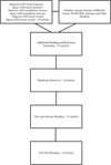
The use of spear gun projectiles is frequent in fishing and water-sports. Their use is uncomplicated, and spear gun injuries are very rare. Most of the cases are related to lack of attention during sports practices [1,2]. Spear injuries may cause severe lesions to the patient including death. This present study aims to describe an unusual case of facial injury associated with a mandible fracture after a spear gun shot. Also, clinical-tomographic features and management, as well as a thorough review of cases found in English language literature from 1983 to 2019 are presented as described in Figure 1. Furthermore, the authors are proposing a treatment guidelines based on data described in the literature.
 |
Fig. 1 Flowchart Diagram of the Up-to-date literature review. |
Case report
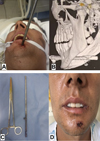
38-years-old man was referred to the Oral and Maxillofacial Surgery Service of a Brazilian Public Hospital, with a history of a penetrating injury on the face caused by an accidental shot from a spear gun, that fired a homemade spear made by the patient himself, that built the spear from steel with three hooks. In the pre-hospital care, the spear was cut and separated from the trident hooks to facilitate the patient's transportation. After initial stabilization of the patient, a computed tomography (CT) was taken to better localization of the spear. The CT demonstrated a large spear impaled in the mandibular symphysis, transfixing the mandibular bone and resting in the sublingual space. An Angio-CT was not necessary due to the lack of important vessels in the anterior mandibular region. The patient was taken to the emergency operation room, and under general anesthesia, with an orotracheal tube as indicated by the anesthesia team due to the situation, the spear was manually removed. There were no concerns on the spear's trident hooks, because the pre-hospital team reported that it was the non-cutting part of the spear that inflicted the patient. With the spear removed, the tube was changed to nasotracheal intubation. The maxillomandibular fixation was made using Erich's arch bars. The mandible was accessed via intraoral vestibular approach and the mandibular fracture was reduced and fixed with two 2.0 non-locking miniplates. After 3 days of hospitalization, the patient was discharged with no functional or sensitive deficits. The postoperative control was uneventful with no signs of infection after 6 months (Fig. 2).
 |
Fig. 2 A) Clinical photography revealing an impacted spear in the anterior aspect of the mandible. B) 3D Reconstruction of the computed tomography revealing the presence of the spear and the depth of the impaction. C) Photography comparing the removed spear with a 18 cm forceps. D) Clinical photography of the patient at the discharge moment, on the third post-operative day. |
Observation and comments
One patient with noncontributory medical, social and cultural records was referred to the Oral and Maxillofacial Surgery Service of a Brazilian Public Hospital. Previous studies of spear gun injuries on the face, published between 1983 and 2019, were researched by means of a detailed investigation of English-language literature across PubMed, by searching the following keywords: “Spear gun injury OR harpoon injury AND face”. All studies that included the complete description of the case, and matched the search strategy were included in this review. Together with this present study, a total of 13 cases were selected. The data from all studies are presented in Table I [1–10].
The main cause for the penetrating injuries caused by spear gun is accident (69.2%), followed by assaults (15.4%) and suicide attempt (15.4%). The majority of cases published in the studies were in male patients (92.4%) with average age of 25.4-years-old. The orbit was the region of the face most affected (41.1%), followed by the Nose (17.6%), frontal region (11.7%) and the infraorbital rim (5.8%), maxilla (5.8%), mouth (5.8%), submental region (5.8%) and mandibular symphysis (5.8%).
The propaedeutic by image examinations are fundamental for cases of penetrating injuries on the face. The X-ray was applied in 84.8% of the cases, CT was used in 69.3% of the cases and angiographic exams in 15.3%.
Surgical removal is mandatory in all cases described in the English-language literature. The direct manual traction of the spear and the removal under direct vision by surgical approach had usage in 46.1% of the cases equally. In one of the cases, the spear was not removed because the clinical condition of the patient. The main complications for these injuries are paresis (23.5%), however, no complications were reported in 46.1% of the cases.
Craniofacial penetrating trauma related to a spear gun injury is extremely rare. It can be life-threatening because the proximity with the neurological structures, and the other vital structures as vessels and the sense organs [1].
It's extremely important to carry out image examinations prior to the surgical procedure to determine the extension of the bone fractures and the injuries in the neurovascular structures. Also, the trajectory of the foreign body is important to planning the surgical procedure defining the point of entry and the point of exit if exist [3]. Conventional radiographic exam is useful in the diagnosis and helps to determine the shape and position of the spears, although the CT scan is essential to demonstrate the exact location and to guide the surgical procedure. The arteriography or angio-CT should be recommended prior to the surgical removal in cases where there is proximity or lesions in major vessels [4].
The surgical procedure should be performed as soon as possible to minimize the risk of infection. In addition, the surgical approach to remove such objects is unique and it should be personalized for each case. The debridement and removal of the object should be done carefully, with the excision of necrotic tissue and surrounding clots [5]. The forced removal of the object must be discouraged, because it is unsuccessful in the majority of cases. Also, large incisions are usually unnecessary and it may cause less favorable esthetic scars and may lead to greater damage to surrounding tissues [6].
Complications associated with this type of injury can be divided in (1) major complications and (2) minor or no complications. Major complications were presented in 30.4% of the cases described in the literature with 1 case of death and 3 cases of blindness after the injuries. The most common complications reported in the literature are paresis representing 23.5% of the cases, partial blindness (15.2%), enucleation off the right eye (7.6%), and even death (7.6%).
At the early postoperative care, the patient should be observed for bleeding and infection. The patient must be advocated to maintain a follow up for at least one year after the improvement of the function or until the rehabilitative follow up of the possible sequels.
Descriptive Statistic of the cases described in the literature, gathering age, gender, etiology, region, image protocol, treatment and complications.
Conclusions
The use of spear gun projectiles is frequent in fishing and water-sports and injuries are very rare. The lesions are severe and it may vary from loss of visual acuity, paresis and even death. An image protocol should be preconized before any surgical procedure, and the CT-Scan is the gold-standard for the recognition of the position, trajectory, neurovascular involvement and to guide the surgical procedure. Angio-CT should be indicated in cases associated with major vessels of the craniofacial region to minimize and/or prevent important bleeding during the surgical procedure. The surgical procedure should be done as soon as possible and the removal of the spear must be done carefully, under direct vision, with or without surgical incisions. Major complications can occur after spear injuries, therefore, the patient must be observed in the postoperative period and should maintain follow up until the end of the rehabilitative process.
Author contribution statement
All authors contributed to the study conception and design. Emergency Care, Surgical Procedure, Material preparation, data collection and analysis were performed by Rodolfo Cesar Gual], Bernardo Barcelos Greco, Alessandro Oliveira de Jesus, Bruna Campos Ribeiro and Marcio Bruno Figueiredo Amaral. The first draft of the manuscript was written by Samuel Macedo Costa and all authors commented on previous versions of the manuscript. All authors read and approved the final manuscript.
Compliance with ethical standards
None of the authors have research funding, nor are they related to companies that may have benefited from hospital or surgical material funding.
The patient was widely informed about the treatment and the conditions for the dissemination of images and publication of the case in a scientific journal. This study was approved by the Ethics committee of the João XXIII hospital under the protocol number n. 924322182.2.0000.5119, and informed written consent forms were obtained from all patients and/or families.
Conflicts of interests
The authors declare that they have no conflicts of interest in relation to this article.
References
- Hefer T, Joachims HZ, Loberman Z, Gdal-on M, Progras Y. Craniofacial speargun injury. Otolaryngol Head Neck Surg 1996;115:553–555. [CrossRef] [PubMed] [Google Scholar]
- Ban LH, Leone M, Visintini P, Blasco V, Antonini F, Kaya JM, et al. Craniocerebral penetrating injury caused by a spear gun through the mouth. J Neurosurg 2008;108:1021–1023. [PubMed] [Google Scholar]
- Gutierrez A, Gil L, Sahuquillo J, Rubio E. Unusual penetrating craniocerebral injury. Surg Neurol 1983;19:541–543. [CrossRef] [PubMed] [Google Scholar]
- Ribeiro ALR, Vasconcellos HG, Pinheiro JAJV. Unusual fishing harpoon injury of the maxillofacial region in a child. Oral Maxillofac Surg 2009;13:243–246. [Google Scholar]
- Lopez F, Martinez-Lage JF, Herrera A, Sanchez-Solis M, Torres P, Palacios MI, et al. Penetrating craniocerebral injury from an under water fishing harpoon. Child's Neri syst 2000;16:117–119. [CrossRef] [Google Scholar]
- Alper M, Totan S, Çankayali R, Songur E. Maxillofacial spear gun accident: report of two cases. J Oral Maxillofac Surg 1997;55:94–97. [CrossRef] [PubMed] [Google Scholar]
- O'neill OR, Gilliland G, Delashaw JB, Purtzer TJ. Transorbital penetrating head injury with a hunting arrow: case report. Surg Neurol 1994;42:494–497. [CrossRef] [PubMed] [Google Scholar]
- Sadda RS. Fish-gun injury of the maxillofacial region. J Oral Maxillofac Surg 1996;54:1132–1135. [CrossRef] [PubMed] [Google Scholar]
- Burnham R, Bhandari R, Holmes S. Diver's Harpoon gun: facial injury caused by an unusual weapon. Br J Oral Maxillofac Surg 2010;482–483. [CrossRef] [PubMed] [Google Scholar]
- Bakohs D, Villeneuve A, Kim S, Lebrun H, Dufour X. Head spear gun injury: an atypical suicide attempt. J Craniofac Surg 2015;26:547–548. [Google Scholar]
All Tables
Descriptive Statistic of the cases described in the literature, gathering age, gender, etiology, region, image protocol, treatment and complications.
All Figures
 |
Fig. 1 Flowchart Diagram of the Up-to-date literature review. |
| In the text | |
 |
Fig. 2 A) Clinical photography revealing an impacted spear in the anterior aspect of the mandible. B) 3D Reconstruction of the computed tomography revealing the presence of the spear and the depth of the impaction. C) Photography comparing the removed spear with a 18 cm forceps. D) Clinical photography of the patient at the discharge moment, on the third post-operative day. |
| In the text | |
Current usage metrics show cumulative count of Article Views (full-text article views including HTML views, PDF and ePub downloads, according to the available data) and Abstracts Views on Vision4Press platform.
Data correspond to usage on the plateform after 2015. The current usage metrics is available 48-96 hours after online publication and is updated daily on week days.
Initial download of the metrics may take a while.


