| Issue |
J Oral Med Oral Surg
Volume 26, Number 3, 2020
|
|
|---|---|---|
| Article Number | 29 | |
| Number of page(s) | 5 | |
| Section | Cas clinique et revue de la littérature / Up-to date review and case report | |
| DOI | https://doi.org/10.1051/mbcb/2020024 | |
| Published online | 26 June 2020 | |
Up-to Date Review And Case Report
Hereditary angioedema type II and dental extraction: case report and literature review
1
Dentistry Department, Tours University Hospital Centre, Tours, France
2
Faculty of Odontology, University of Clermont Auvergne, Clermont-Ferrand, France
3
Dermatology Department, Tours University Hospital Centre, Tours, France
4
Faculty of Odontology of Nantes, University of Nantes, Nantes, France
* Correspondence: This email address is being protected from spambots. You need JavaScript enabled to view it.
Received:
15
April
2020
Accepted:
26
May
2020
Abstract
Introduction: The hereditary angioedema of type II (HAE type II) is a disease which, in the case of invasive acts such as dental extractions, can trigger potentially fatal laryngeal edema. Observation: A 64-year-old man presented to the hospital to perform dental extractions. The anamnesis revealed a history of hereditary angioedema of type II. The National Reference Center for Angioedema (CREAK) recommended a prophylactic protocol before performing this traumatic dental procedure in order to reduce the risk of post-operative edema. Discussion: The HAE type II is under- or misdiagnosed. The literature reports cases of death related to dental procedures in patients unaware of being a carrier of the disease. Conclusion: In case of certain evocative signs (recurrent edema, abdominal pain, family history, etc.), the odontologist must play an active role in the early detection of HAE type II.
Key words: angioedema / hereditary / dental
© The authors, 2020
 This is an Open Access article distributed under the terms of the Creative Commons Attribution License (https://creativecommons.org/licenses/by/4.0), which permits unrestricted use, distribution, and reproduction in any medium, provided the original work is properly cited.
Introduction
The hereditary angioedema (HAE) type II is a rare and life-threatening genetic disorder that is often under- or misdiagnosed [1]. It is caused by mutations in the SERPING 1 gene, which causes a qualitative defect in the C1 esterase inhibitor (C1-Inh), leading to an abrupt and localized release of bradykinin, a peptide with vasodilating properties. This leads to an increased vascular permeability which can lead to edema of the face, limbs, upper airways or digestive tract.
The edema is sudden, subcutaneous and/or submucosal, circumscribed, white, non-pruritic and disappears without sequelae in 2–5 days [1]. The appearance of HAE is spontaneous or caused by local trauma such as oral care, by taking certain medications or by emotional stress [2]. The first signs of HAE can occur at any age but most often begin in childhood [3].
Facial edema (e.g. following dental treatment) is at high risk for laryngeal damage, with a risk of death by asphyxiation in the absence of specific treatment. It is therefore essential, before treating any new patient, to collect a complete medical history and apply a specific protocol.
Observation
The reported case is that of a 64-year-old man referred by his dental surgeon to the dentistry department of his local university hospital for dental extractions. In this patient, a diagnosis of HAE type II had been made in 2009 by the CREAK. He also suffered from lumbar osteoarthritis and dyslipidemia. The patient was treated with icatibant (FIRAZYR®), 30 mg in case of an attack.
The first manifestations of the disease appeared within his childhood (5–6 years), as edemas with multiple localizations: abdomen, hand, foot, face. Despite a family history, the patient was diagnosed at 54 years of age. With age, the attacks seem to diminish in frequency and intensity. Spontaneous edema occurs, approximately every 3 weeks, touching a single finger and disappearing during the day. In 2020, endodontic treatment without a prophylactic protocol resulted in facial edema (without laryngeal involvement) that disappeared after 2–3 days. In 2017, dental extractions were performed and in 2018 implants were placed with a prophylactic protocol 6 hours before the intervention with a C1Inh concentrate (BERINERT®), 20 U/kg. In both cases, there were no post-operative complications.
At the intraoral clinical examination, the patient complained of discomfort in the maxillary teeth. The gum next to the teeth n°25-26-27 was inflammatory. Two temporary bridges, from tooth n°13 to tooth n°16 and from tooth n°22 to tooth n°27 were present and mobile (Fig. 1a). The survey revealed periodontal pockets in teeth n°16-22-25-26-27. A recurrence of decay lesions was observed in these same teeth. In the mandible, the teeth were healthy and asymptomatic. The orthopantomogram showed periradicular radiolucency in teeth n°16-22-25-26-27 and horizontal alveolisis reaching 1/3 of the root height (Fig. 1b).
After multidisciplinary consultation, an appointment was scheduled to perform avulsions of teeth n°16-22-25-26-27 (Fig. 1c). In the context of an hospitalisation, the following protocol was set up following the recommendations of the CREAK: a C1Inh concentrate injection (BERINERT®), 20 U/kg, in the 6 hours preceding the procedure to be completed in the event of an attack with icatibant (FIRAZYR®), 30 mg. During the procedure, additional precautions were taken in order to limit the risk of post-operative edema: the choice of a para-apical local anesthetic (articaine with epinephrine 1/200000) was made and injected slowly to prevent any risk of trauma.
Likewise, multiple-rooted teeth extractions were performed with root separation, and flapless. (Fig. 1c).
At the end of the procedure, a transitional partial denture was placed (Fig. 1d). Before installing the device, the surface condition was checked to avoid any mucosal trauma. Post-operative advices have been clarified. A stage I analgesic and an antiseptic solution were ordered. The patient remained hospitalized for the night, for observation. No laryngeal edema occurred.
At the post-operative appointment (J14), healing was satisfactory (Fig. 1e). The sutures have been removed and the transitional removable device was readjusted at this time (Fig. 1f) to avoid any trauma to the mucosa.
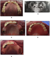 |
Fig. 1 Iconographies of the clinical case. (a) Intraoral photography in preoperative. (b) Orthopantomogram. (c) Maxillary occlusal view in peroperative. (d) Removal partial denture. (e) Postoperative view. |
Discussion
HAE of type II is a rare disease [4], which can lead to life-threatening edema of the limbs and face. In order to enrich the presentation of this case, a review of the literature on the PubMed search engine was conducted from 2002 to 2019 with the following search equation: [hereditary] AND [angioedema] AND [dental]. 12 articles have been retained among 78 articles initially selected (Fig. 2). Articles were excluded if it was clear from the title of the article or abstract that the topic was not relevant or did not meet the criteria of the literature review (articles relating to cases of HAE of type II related to dental care).
The main causes of the appearance of HAE reported in the literature are dental extractions, taking certain medications, stress, pulp devitalization and taking dental impressions (Table I). Different care protocols are reported in relation to the performance of the various acts of odontostomatology.
Dental extractions appear to be the most common trigger for HAE type II, since 8 cases have been reported in the literature [6,9,10]. Among these 8 cases, two had a fatal outcome [6,9]. In both cases, the patient did not know that he was a carrier of HAE. It should be noted that in most cases the edema arrives late after the operation, sometimes more than 2 days later, which underlines the importance of post-operative follow-up [10]. In this case, the patient should be warned that edema may appear even several days after the operation. The patient must be attentive to any respiratory difficulties in order to act quickly. Dysphagia, a weak or inaudible voice can also be the prodromes of a future laryngeal attack.
Certain drug treatments also appear to be a trigger. Hammond et al. report a case of a patient with HAE type II after taking ibuprofen and codeine [5].
Psychological stress may trigger edema [4]. For this reason, prior to tooth extraction in a patient with HAE, Rosa et al. describe the use of conscious sedation to reduce anxiety and thus pain perception to help prevent post-operative angioedema [4].
Baliga et al. described a case of HAE type II following pulp devitalization. In this case, the patient was unaware that she had this disease and no family history had been reported [7]. More marginally, a case of HAE was described following dental impression taking [8].
Short-term prophylaxis consisting of the injection of C1-Inh within 6 hours before the procedure does not completely avoid the risk of postoperative edema, which is why the use of icatibant (FIRAZYR®) or the concentrate injection of C1-Inh (20 U/kg) may be necessary postoperatively [12].
Early diagnosis of this disease is essential [11] although the lack of specificity of symptoms complicates the diagnosis [1]. In this context, most incidents during dental care have occurred in patients who were unaware of being a carrier of this disease [13,6,5,9]. HAE type II should be mentioned in the case of any clinical signs suggestive of the disease: isolated non-erythematous and non-pruritic angioedema, unexplained recurrent abdominal pain, recurrent oral edema or a family context [11]. However, only the quantitative and functional C1-INH assays and the C4 Complement Compound assay can certify the diagnosis. A concentration of C1-INH lowered on two samples allows to confirm the diagnosis. The treatment of the pathology takes place in specialized centers, the CREAKs [16].
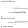 |
Fig. 2 Flow chart. |
Summary of selected articles.
Conclusion
This report shows the importance of early diagnosis of HAE type II, allowing effective multidisciplinary care and control of a risk management of post-operative edema. Mobilization of practitioners to know and suspect this disease must be important in view of the fatal nature of the attacks. In the case of recurrent and transient edema, type II HAE should be mentioned and a consultation at the CREAK reference center should be considered. Studies are underway to improve the early diagnosis. The genotype-based approach should eventually allow earlier detection of this type of pathology.
Conflicts of interests
The authors declare that they have no conflicts of interest in relation to the publication of this article.
References
- Bodian DL, Vilboux T, Hauser NS. Genotype-first analysis of a generally healthy population cohort supports genetic testing for diagnosis of hereditary angioedema of unknown cause. Allergy Asthma Clin Immunol 2019;15:32. [CrossRef] [PubMed] [Google Scholar]
- Longhurst HJ, Bork K. Hereditary angioedema: an update on causes, manifestations andtreatment. Br J Hosp Med (Lond) 2019;80:391–398. [CrossRef] [PubMed] [Google Scholar]
- Dermesropian A. Épidémiologie descriptive de l'angioedème à bradykinines chez l'enfant sur le territoire français. 2015;71. [Google Scholar]
- Rosa A, Miranda M, Franco R, Guarino MG, Barlattani A, Bollero P. Experimental protocol of dental procedures In patients with hereditary angioedema: the role of anxiety and the use of nitrogen oxide. Oral Implantol (Rome) 2016;9:49–53. [PubMed] [Google Scholar]
- Hammond D, Olaore S, Salker M, Gallaway E. Catheter-based local analgesia for the fractured mandible in a patient with a history of hereditary angioedema. J Surg Case Rep 2019;2019:rjz126. [CrossRef] [PubMed] [Google Scholar]
- Rice S, Cochrane TJ, Millwaters M, Ali NT. Emergency management of upper airway angio-oedema after routine dental extraction in a patient with C1 esterase deficiency. Br J Oral Maxillofac Surg 2008;46:394–396. [CrossRef] [PubMed] [Google Scholar]
- Baliga M, Ramanathan A, Bhambar RS. Angioedema triggered by pulp extirpation − a case report. Oral Maxillofac Surg 2011;15: 253–255. [Google Scholar]
- Aziz SR, Tin P. Spontaneous angioedema of oral cavity after dental impressions. N Y State Dent J 2002;68:42–45. [Google Scholar]
- Forrest A, Milne N, Soon A. Hereditary angioedema: death after a dental extraction. Aust Dent J 2017;62:107–110. [CrossRef] [PubMed] [Google Scholar]
- Bork K, Barnstedt S-E. Laryngeal edema and death from asphyxiation after tooth extraction in four patients with hereditary angioedema. J Am Dent Assoc 2003;134:1088–1094. [CrossRef] [PubMed] [Google Scholar]
- Floccard B, Crozon J, Rimmelé T, Vulliez A, Coppere B, Chamouard V, et al. [Management of bradykinin-mediated angioedema]. Ann Fr Anesth Reanim 2011;30:578–588. [CrossRef] [PubMed] [Google Scholar]
- Williams AH, Craig TJ. Perioperative management for patients with hereditary angioedema. Allergy Rhinol (Providence) 2015;6:50–55. [CrossRef] [PubMed] [Google Scholar]
- Hosokawa R, Tsukamoto M, Nagano S, Yokoyama T. Anesthetic management of a patient with hereditary angioedema for oral surgery. Anesth Prog 2019;66(1):30–32. [Google Scholar]
- Waldon K, Barber SK, Spencer RJ. Orthodontic treatment for a patient with hereditary angiodema: a case report. Int J Paediatr Dent 2015;25:229–232. [CrossRef] [PubMed] [Google Scholar]
- Papamanthos M, Matiakis A, Tsirevelou P, Kolokotronis A, Skoulakis H. Hereditary angioedema: three cases report, members of the same family. J Oral Maxillofac Res. 2010;1:e9. [CrossRef] [PubMed] [Google Scholar]
- Bouillet L, Defendi F, Hardy G, Cesbron JY, Boccon-Gibod I, Deroux A, et al. Diagnostic biologique des angioedèmes bradykiniques: les recommandations du CREAK. La Presse Médicale 2019;48:55–62. [CrossRef] [Google Scholar]
All Tables
All Figures
|
Fig. 1 Iconographies of the clinical case. (a) Intraoral photography in preoperative. (b) Orthopantomogram. (c) Maxillary occlusal view in peroperative. (d) Removal partial denture. (e) Postoperative view. |
| In the text | |
|
Fig. 2 Flow chart. |
| In the text | |
Current usage metrics show cumulative count of Article Views (full-text article views including HTML views, PDF and ePub downloads, according to the available data) and Abstracts Views on Vision4Press platform.
Data correspond to usage on the plateform after 2015. The current usage metrics is available 48-96 hours after online publication and is updated daily on week days.
Initial download of the metrics may take a while.