Fig. 4
Download original image
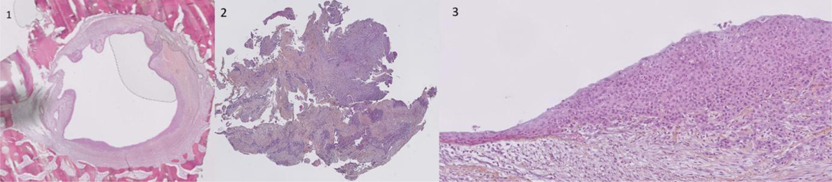
Three histological sections from the initial biopsy: No 1 (left) — Well-circumscribed cystic lesion located within the bone. H&E, original magnification × 20. No 2 (center) — Initial biopsy showing a well-differentiated invasive squamous cell carcinoma with a lobulated architecture. H&E, original magnification × 40. No 3 (right) — Same cystic lesion squamous epithelium transitioning from a normal appearance (left) to carcinoma in situ and then to invasive carcinoma with stromal invasion. H&E, original magnification × 200.
Current usage metrics show cumulative count of Article Views (full-text article views including HTML views, PDF and ePub downloads, according to the available data) and Abstracts Views on Vision4Press platform.
Data correspond to usage on the plateform after 2015. The current usage metrics is available 48-96 hours after online publication and is updated daily on week days.
Initial download of the metrics may take a while.