| Issue |
J Oral Med Oral Surg
Volume 30, Number 2, 2024
|
|
|---|---|---|
| Article Number | 18 | |
| Number of page(s) | 7 | |
| DOI | https://doi.org/10.1051/mbcb/2023038 | |
| Published online | 04 October 2024 | |
Original Research Article
Oro-antral communication closure using collagenated porcine cortical lamina: A retrospective study of 34 cases
1
Department of oral and maxillo-facial surgery, Hospital Novo, Pontoise
2
Department of oral surgery, Hospital Pitié Salpétrière, Paris
3
Department of Medical and Surgical Intensive Care, Hospital Novo, Pontoise
4
Department of Oral and Maxillo-facial surgery, Hospital Novo, Pontoise
* Correspondence: This email address is being protected from spambots. You need JavaScript enabled to view it.
Received:
7
August
2023
Accepted:
30
October
2023
Abstract
Introduction: Oro-antral communications (OAC) is a common complication in oral surgery. Many surgical techniques have been described, but traditional closure techniques have limitations, leading to the exploration of alternative approaches using resorbable biomaterials. This study aimed to assess the success of repairing OAC larger than 5 mm using collagenated porcine cortical lamina. Materials and methods: This retrospective study included 34 cases of OAC larger than 5 mm who underwent surgical repair using a porcine-derived collagenated cortico-cancellous plate, (Lamina Curve®). The median patient age was 46 yr. The study cohort consisted of 12 females and 22 males. The median follow-up time was 54 days. The primary outcome was the presence of complete mucosal closure, assessed at the 4th week postoperatively. Secondary outcomes included adverse events and stitching disunion. Results: Success rate was 97%. 9 patients (26.5% [12.1 ; 44]) had complications : 5 stitch disunions, 1 failure, 1 epistaxis, 1 infection and 1 nerve paresis. Of the 5 patients with stitch disunions (14.7% [4.12 ; 30.2]), all patients had complete mucosal closure at the primary outcome endpoint. Conclusion: Cortical lamina shows promising results in OAC repair. Success rates compare favorably with traditional flap-based methods. Further research is needed to validate these findings.
Key words: Oro-antral communication / sinusitis / heterologous bio material / oral surgery
© The authors, 2024
 This is an Open Access article distributed under the terms of the Creative Commons Attribution License (https://creativecommons.org/licenses/by/4.0), which permits unrestricted use, distribution, and reproduction in any medium, provided the original work is properly cited.
Introduction
Oroantral communication (OAC) is an abnormal communicating path that forms between the maxillary sinus and oral cavity. It most often follows the enucleation of a cyst in the posterior maxillary region, trauma [1] or the extraction of posterior maxillary teeth whose root anatomy is very close to the sinus (on average between 1 mm and 7 mm) [2]. A fistula larger than 5 mm does not tend to collapse spontaneously [3] and requires surgical closure. When an OAC develops, the presence of maxillary sinusitis, the formation of epithelial tissue along the fistula tract, the occurrence of a dental apical abscess, inflammation of the bone (osteitis) or bone infection (osteomyelitis) near the communication site, the presence of dental cysts, foreign bodies, or tumors will impede spontaneous healing and lead to the formation of a persistent fistula [4]. Therefore, it is crucial to address the pathological conditions affecting the maxillary sinus for the successful treatment of OAC. Proper drainage and sufficient ventilation of the sinus need to be achieved. Moreover, foreign bodies, infected and degenerated polypoid mucosa, as well as infected bone, should be promptly removed, and the defect should be closed through surgical means [5]. Many OAC closure techniques have been developed but two techniques are widely used: the vestibular flap (VF) and the buccal fat pad flap using Bichat's fat pad (BFP). The vestibular flap described by Rehrmann in 1936 is the reference surgery for minor OAC [6]. The use of the Bichat fat pad for OAC closure was proposed by Egyedi in 1974 [7] and is generally used for closure of OAC larger than 5 mm [5,8]. The closure of OAC by palatal flap (PF) is less used due to the presence of the palatal artery and the need to leave part of the palate bare during healing [5]. These very simple techniques have a relatively high success rate: between 89.8% [5] and 93% [9] for the vestibular flap and almost 100% [10–12] for the Bichat fat pad. The palatal flap has a success rate of 76% [13]. However, the use of flap-based method, in particular the BFP does induce very little to no new bone formation in the sinus floor [14]. Additionally, retracted and collapsed soft tissues from the vestibular region can lead to scarring and changes in the mucosal topography inducing difficulties in further prosthetic or implant rehabilitation [15]. Furthermore, graft contraction can cause limitation in mouth opening, which is one of the main disadvantages of BFP [16]. The presence of these constrains, combined with the occurrence of a persistent oroantral communication (OAC) even after attempted closure using vestibular or palatal flaps, and the potential risks of damaging the facial nerve during Bichat fat pad dissection, have prompted the exploration of alternative closure approach involving heterologous resorbable biomaterial. Besides closure of the opening, these materials may positively affect the surrounding hard tissues as the use of resorbable heterologous cortical lamina has been reported to reduce alveolar ridge resorption [17] and had been used for crestal ridges reconstruction [18].
The purpose of this study is to assess the success in the repair of OAC larger than 5 mm using resorbable biomaterials of porcine origin, in particular, cortical lamina.
Materials and methods
Study design
This retrospective study included patients over 18 yr old with a diagnosed OAC larger than 5 mm, who underwent surgical repair using the specified Lamina Curve plate between 2016 and 2023. Patients with a follow-up time of less than 1 month after surgery and those unable to be informed about the study or who did not provide consent were excluded. A total of 35 patients were initially screened. However, one patient with an OAC size smaller than 5 mm and two patients who were unable to provide informed consent were excluded from the study. After exclusions, the medical records of 33 patients were included in the analysis. Among these patients, one had bilateral OAC, resulting in a total of 34 OAC cases treated with a Lamina Curve plate. Patient records were examined to collect relevant information, including patient age, gender, OAC size, tooth location, follow-up time, and postoperative adverse events. The median age of the patients was 46 yr, with an interquartile range (IQR) from 38 to 66 yr (Tab. I). The study cohort consisted of 12 female participants (35%) and 22 male participants (65%) (Tab. I). The average number of days for the last follow-up was 54 (median, interquartile range: 41–204). The primary outcome was the presence of complete mucosal closure four weeks after the surgery. (median day of evaluation: 35, IQR: 32.3 − 38.0) (Tab. I). Secondary outcome measures included the presence of adverse events and stitching disunion. The data collected from the medical records were analyzed descriptively. The interpretation of the results was performed using median, interquartile range and percentages by comparing the collected data. To facilitate a better assessment and analysis, the results were organized into tables. Statistical analysis was performed by P. Michel M.D. (Medical and Surgical Intensive Care Dep., Hospital Novo, Pontoise). This study was conducted in accordance with the ethical guidelines set forth by the Declaration of Helsinki. The research protocol was registered on ClinicalTrial.gov before the data collection process (Identifier: NCT05910073). Patient confidentiality and anonymity were ensured throughout the study, and all patient identifiers were removed during data analysis to maintain privacy. Information notes were sent to patients. If no reply was received within 15 days, the patient was deemed not to have objected.
Patients caracteristics
Biomaterials and fixation devices
The biomaterial used consisted of a porcine-derived collagenated cortico-cancellous shield (Lamina® Curve 35 × 35 × 1 mm, OsteoBiol®, Tecnoss Srl). This membrane is a soft cortical lamina derived from cortical porcine bone, with a plastic consistency. It can be shaped each time with sterile scissors to reach the desired size, hydrated in sterile physiological solution until the desired plasticity is acquired, and adapted to completely cover the OAC site. 5 mm titanium osteosynthesis screws (Leibinger, Stryker®) were used to stabilize the plate.
Surgical procedure
After disinfection with a Betadine® solution and infiltration with a 0.2% adrenaline xylocaine anesthetic, a full thickness vestibular flap was lifted with vertical mesial discharge 1 to 2 teeth in front of the fistula (Figs. 1, 2A, and 2B). A sinusotomy was performed with an abundant Betadine® physiological saline solution wash. The Lamina® plate was hydrated with physiological serum until it was sufficiently flexible. It was then shaped and trimmed according to the anatomical characteristics and secured with vestibular and/or palatal osteosynthesis screws (Fig. 2C). The vestibular and palatal flaps were elongated in order to obtain a tight closure on a tensionless flap. The antibiotic therapy consisted of amoxicillin and clavulanic acid (Augmentin®) 3 g daily for 7 days. In case of penicillin allergy, patients were given clindamycin 1200 mg and 1500 mg metronidazole daily for 7 days. Analgesic treatment was administered depending on the pain. A first check was carried out on day 7 postoperatively to asses for the presence of any adverse events or stitching disunion. Then on day 30 full mucosal closure was clinically evaluated.
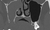 |
Fig. 1 Pre operative TDM showing the oro-antral communication and the opacification of the left maxillary sinus. |
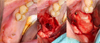 |
Fig. 2 (A) Buccal views of the oro-antral fistula, (B) buccal and palatal flaps, (C) fixation of the membrane. |
Results
The median size of the OAC was 10 mm (interquartile range 9–10 mm). 23 (68%) (Tab. I) patients had chronic sinusitis and 16 (47%) patients where smokers (Tab. I). 33 patients out of the 34 had a mucosal closure at the primary outcome endpoint making for a success rate of 97% (Tab. I). The only patient who did not have complete mucosal closure suffered from osteochimionecrosis of the jaw due to anti-resorptive bone therapy. 9 patients (26.5% [12.1; 44]) had a complication : 5 stitch disunions, 1 failure, 1 epistaxis, 1 infection and 1 infra-orbital nerve paresis (Tab. II). Of the 5 patients with stitch disunions (14.7% [4.12; 30.2]), all patients had complete mucosal closure at the primary outcome endpoint (Tab. III).
Complications
Stitch disunion.
Discussion
The successful closure of Oroantral Communications (OAC) remains a critical aspect of patient care, as untreated OACs can lead to various complications, including sinusitis, infection, and osteomyelitis. In this study, we aimed to assess the effectiveness of using resorbable biomaterials of porcine origin, specifically cortical lamina, for the repair of OACs larger than 5 mm.
The results of this study demonstrated a high success rate of 97% in achieving complete mucosal closure at the primary outcome endpoint. This finding is encouraging and suggests that the use of resorbable cortical lamina is a viable alternative for OAC repair, especially for larger defects. The success of this technique can be attributed to the high biocompatibility of the membrane. Indeed, histologic analysis of the cortical lamina after six months revealed nearly complete resorption and substitution by newly formed connective tissue, indicating a certain porosity that allows vascular cells' ingrowth and new vessel formation [19]. This finding suggests its potential as a biologic and stable barrier during the healing process. The slow resorption rate of the cortical lamina may be beneficial, as it provides a prolonged period for osteoconductive properties to promote bone formation and soft tissue healing [20]. The finding that all five patients with stitch disunions achieved complete mucosal closure at the primary outcome endpoint is interesting and may be attributed to the resorbable nature of the Lamina Curve plate. The resorbable Lamina Curve may act as a scaffold for the development of granulation tissue and subsequent keratinized mucosa and as authors have noted may promote a second intention healing process in case of exposure [19]. It is worth noting that the patient who did not achieve complete mucosal closure suffered from osteochimionecrosis of the jaw due to anti-resorptive bone therapy. This finding highlights the importance of considering individual patient factors and medical history when choosing the appropriate treatment approach for OAC closure. Additionally, the presence of complications in 26.5% of patients, including stitch disunions, epistaxis, infection, and infra-orbital nerve paresis, indicates that while the overall success rate is high, careful consideration of potential risks and complications is essential when using this technique. Comparing our findings with previous studies using traditional closure techniques such as vestibular flap (VF) and buccal fat pad flap (BFP), the success rate observed in this study with resorbable cortical lamina is comparable or even superior to the reported success rates for VF (89.8% to 93%) and BFP (almost 100%). This suggests that the use of resorbable biomaterials offers a promising alternative to conventional flap-based methods for OAC repair, especially considering that flap-based approaches may have limitations such as limited bone formation in the sinus floor and scarring of soft tissues in the vestibular region. From a practical point of view, the curved and flexible nature of this plate made the procedure relatively easy as the membrane can fit the contours of the bone defect and slide under the vestibular and palatal flaps. The main challenge of this procedure is to obtain a tensionless flap.
To the best of our knowledge, no previous study has explored the use of resorbable biomaterials of porcine origin for OAC closure. Kapustecki et al. [21] used autogenous bone graft and plasma rich fibrin (PRF) as a membrane in 20 patients, observing successful closure of the OAC in all cases. Their study focused on normal bone regeneration at the OAC site and preparation of alveolar bone for new implants and prosthetic solutions. On the other hand, Gacic et al. [15] explored the effectiveness of hemostatic gauze and PLGA-coated ß-TCP for OAC closure. Their minimally invasive methods showed similar effectiveness to the well-established buccal flap approach, with lower pain intensities and reduced swelling in patients treated with PLGA-coated ß-TCP granules. These studies have contributed valuable insights to the field of OAC closure techniques. In comparison, our study highlighted the potential advantages of using the resorbable Lamina Curve plate, including its ability to promote second intention healing with keratinized mucosa in cases of exposure, which may offer benefits in terms of enhanced healing and reduced complications in OAC repair.
While the findings of this study are encouraging, several limitations should be acknowledged. First, the study's retrospective nature may introduce selection bias and limit the control over confounding variables. The relatively small sample size might affect the generalizability of the results. Moreover, the study's follow-up period varied between 41 days and 6 months, possibly limiting its ability to fully capture longer-term outcomes. It's important to note that the primary focus of this innovative use of the cortical lamina was to evaluate OAC closure from a clinical perspective. Consequently, no imaging techniques were employed to assess potential bone formation. To address this, future investigations utilizing advanced imaging methods such as Cone Beam Computed Tomography (CBCT) could provide valuable insights and quantitative data regarding bone formation. The short-term nature of this study restricted our capacity to comprehensively analyze extended-term results. However, it's worth mentioning that histologic analysis indicated a likely resorption of the cortical lamina, with potential substitution by new bone at the 6-month mark.
Further research with larger sample sizes, extended follow-up periods, and comprehensive imaging assessments is needed to validate and expand on the current findings.
Conclusion
This study provides valuable insights into the successful repair of OACs larger than 5 mm using resorbable biomaterials of porcine origin, particularly cortical lamina. The high success rate observed in achieving complete mucosal closure and the potential positive effects on surrounding hard tissues make this approach a promising option for OAC repair. The results of this study contribute to the existing knowledge in the field and support the exploration of alternative closure approaches involving resorbable biomaterials for OAC treatment. Further prospective studies with longer follow-up periods are necessary to corroborate these findings and assess the long-term outcomes of this novel technique.
Funding
This research received no external funding.
Conflicts of interest
The authors declare that there are no conflicts of interest regarding the publication of this paper.
Data availability statement
The data that support the findings of this study are available from the corresponding author upon reasonable request.
References
- Eppley B, Sclaroff A. Oro-nasal fistula secondary to maxillary augmentation. Int J Oral Surg. 1984;13:535–538. [CrossRef] [Google Scholar]
- Skoglund LA, Pedersen SS, Holst E. Surgical management of 85 perforations to the maxillary sinus. Int J Oral Surg. 1983;12:1–5. [CrossRef] [Google Scholar]
- Awang MN. Closure of oroantral fistula. Int J Oral Maxillofac Surg. 1988;17:110–115. [CrossRef] [Google Scholar]
- Yalçın S, Öncü B, Emes Y, Atalay B, Aktaş İ. Surgical treatment of oroantral fistulas: a clinical study of 23 cases. J Oral Maxillofac Surg. 2011;69:333–339. [CrossRef] [Google Scholar]
- Gheisari R, Hosein Zadeh H, Tavanafar S. Oro -antral fistula repair with different surgical methods: a retrospective analysis of 147 cases. J Dent. 2019;20:107–112. [Google Scholar]
- Rehrmann A. Eine methode zur schliessung von kieferhohlenperforationen. Dtsch Zahnarztl Wschr. 1936;39:1136. [Google Scholar]
- Egyedi P. Utilization of the buccal fat pad for closure of oro-antral and/or oro-nasal communications. J Maxillofac Surg. 1977;5:241–244. [CrossRef] [Google Scholar]
- Abuabara A, Cortez ALV, Passeri LA, de Moraes M, Moreira RWF. Evaluation of different treatments for oroantral/oronasal communications: experience of 112 cases. Int J Oral Maxillofac Surg. 2006;35:155–158. [CrossRef] [Google Scholar]
- Killey HC, Kay LW. Observations based on the surgical closure of 362 oro-antral fistulas. Int Surg. 1972;57:545–549. [Google Scholar]
- El-Hakim IE, El-Fakharany AM. The use of the pedicled buccal fat pad (BFP) and palatal rotating flaps in closure of oroantral communication and palatal defects. J Laryngol Otol. 1999;113:834–838. [CrossRef] [PubMed] [Google Scholar]
- Stajčić Z. The buccal fat pad in the closure of oro-antral communications: a study of 56 cases. J Cranio-Maxillofac Surg. 1992;20:193–197. [CrossRef] [Google Scholar]
- Baumann A, Ewers R. Application of the buccal fat pad in oral reconstruction. J Oral Maxillofac Surg Off J Am Assoc Oral Maxillofac Surg. 2000;58:389–392; discussion 392–393. [CrossRef] [Google Scholar]
- Lee J-J, Kok S-H, Chang H-H, Yang P-J, Hahn L-J, Kuo Y-S. Repair of oroantral communications in the third molar region by random palatal flap. Int J Oral Maxillofac Surg. 2002;31:677–680. [CrossRef] [Google Scholar]
- Visscher SH, van Minnen B, Bos RRM. Closure of oroantral communications: a review of the literature. J Oral Maxillofac Surg Off J Am Assoc Oral Maxillofac Surg. 2010;68:1384–1391. [CrossRef] [Google Scholar]
- B G, L T, V K, V D, L S-S, R D, A M. The closure of oroantral communications with resorbable PLGA-coated beta-TCP root analogs, hemostatic gauze, or buccal flaps: a prospective study. Oral Surg Oral Med Oral Pathol Oral Radiol Endod. 2009;108:844–850. https://doi.org/10.1016/j.tripleo.2009.07.026 [Google Scholar]
- Chien C-Y, Hwang C-F, Chuang H-C, Jeng S-F, Su C-Y. Comparison of radial forearm free flap, pedicled buccal fat pad flap and split-thickness skin graft in reconstruction of buccal mucosal defect. Oral Oncol. 2005;41:694–697. [CrossRef] [Google Scholar]
- Festa VM, Addabbo F, Laino L, Femiano F, Rullo R. Porcine-derived xenograft combined with a soft cortical membrane versus extraction alone for implant site development: a clinical study in humans: alveolar ridge preservation vs extraction alone. Clin Implant Dent Relat Res. 2011;707–713. 10.1111/j. 1708–8208. 2011.00398.x [Google Scholar]
- Lopez MA, Andreasi Bassi M, Confalone L, Carinci F, Ormianer Z, Lauritano D. The use of resorbable cortical lamina and micronized collagenated bone in the regeneration of atrophic crestal ridges: a surgical technique. Case series. J Biol Regul Homeost Agents. 2016;30:81–85. [Google Scholar]
- Rossi R, Rancitelli D, Poli PP, Polo MRD, Nannmark U, Maiorana C. The use of a collagenated porcine cortical lamina in the reconstruction of alveolar ridge defects. A clinical and histological study. Minerva Stomatol. 2016;65:257–268. [Google Scholar]
- Pagliani L, Andersson P, Lanza M, Nappo A, Verrocchi D, Volpe S, Sennerby L. A collagenated porcine bone substitute for augmentation at Neoss implant sites: a prospective 1-year multicenter case series study with histology. Clin Implant Dent Relat Res. 2012;14:746–758. [CrossRef] [PubMed] [Google Scholar]
- Kapustecki M, Niedzielska I, Borgiel-Marek H, Różanowski B. Alternative method to treat oroantral communication and fistula with autogenous bone graft and platelet rich fibrin. Med Oral Patol Oral Cirugia Bucal. 2016;21:e608–e613. [Google Scholar]
All Tables
All Figures
|
Fig. 1 Pre operative TDM showing the oro-antral communication and the opacification of the left maxillary sinus. |
| In the text | |
|
Fig. 2 (A) Buccal views of the oro-antral fistula, (B) buccal and palatal flaps, (C) fixation of the membrane. |
| In the text | |
Current usage metrics show cumulative count of Article Views (full-text article views including HTML views, PDF and ePub downloads, according to the available data) and Abstracts Views on Vision4Press platform.
Data correspond to usage on the plateform after 2015. The current usage metrics is available 48-96 hours after online publication and is updated daily on week days.
Initial download of the metrics may take a while.