| Issue |
J Oral Med Oral Surg
Volume 27, Number 1, 2021
|
|
|---|---|---|
| Article Number | 2 | |
| Number of page(s) | 2 | |
| DOI | https://doi.org/10.1051/mbcb/2020041 | |
| Published online | 22 September 2020 | |
Short Case Report
Diagnostic difficulty of a mandibular manifestation of diffuse large B-cell lymphoma: a case report
1
University of Paris, Paris, France
2
Department of Oral Medicine and Oral Surgery, Bretonneau Hospital, AP-HP, Paris, France
3
Department of Oral Medicine and Oral Surgery, Bichat-Claude Bernard Hospital, AP-HP, Paris, France
4
Department of Otorhinolaryngology, Bichat-Claude Bernard Hospital, AP-HP, Paris, France
* Correspondence: This email address is being protected from spambots. You need JavaScript enabled to view it.
Received:
20
April
2020
Accepted:
23
August
2020
Observation
An 87-year-old patient was referred by his dentist for multiple tooth extractions following pain on the lower right jaw (quadrant 4) and one month of unsuccessful antibiotics treatment. The patient had a medical history of slow progressing myeloma for more than two years with no treatment required, chronic kidney failure and atrial fibrillation.
Extraoral examination revealed a right submandibular lymphadenopathy.
Intraoral examination revealed severe periodontitis characterized by poor oral hygiene, tooth mobility especially in the molars sectors (score 3 and 4 of Muhlemann) and necrotic gum on quadrant 4. The patient also reported spontaneous loss of teeth.
The orthopantomogram (OPT) showed a radiographic bone loss extending to mid third of the roots and beyond.
The patient was treated by apixaban and bisoprolol. He had never received biphosphonate or RANK/RANKL inhibitor.
The main complaint was pain in quadrant 4. Teeth 45 46 47 were hopeless and their extraction was planned.
Extractions were performed with no premedication. After local anesthesia, analgesia was hard to obtain. The extractions revealed a necrotic aspect of the underlying bone and a biopsy was performed looking for osteitis or malignant disease (Fig. 1). Medication-related jaw osteonecrosis was excluded after exhaustive anamnesis. A CBCT was performed to evaluate the extension of the bone necrosis. At this stage it was not informative (Fig. 2).
At follow-up appointment two weeks later, a worsening of the wound was observed, with pain preventing alimentation. A new CBCT was performed and showed small radiolucent lesions in the cortical bone surrounding the extraction site (Fig. 2). Surprisingly, histological result was in favor of actinomycosis.
Considering the worsening of the patient's physical condition (asthenia and pain), necrosis extension, and inaccordance with histological results, the patient was referred to the otorhinolaryngology department. New biopsies with specific markers (CD20+, CD10−, BCL6+, PAX5−, BCL2−, Ki67 = 90%) allowed diagnosis of diffuse large B-cell lymphoma (DLBCL). The tumor extent assessment by CT revealed a pleural effusion linked to the lymphoma.
The patient was then referred to the hematology department to start a treatment with rituximab and bendamustine. Unfortunately, due to the worsening of the patient's physical condition, the second cure could not be delivered. The patient was transferred into palliative care afterwards.
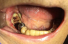 |
Fig. 1 Post-extraction intraoral view. |
 |
Fig. 2 CBCT imaging at the day of the extraction and at the follow-up appointment at 2 weeks. |
Commentary
Lymphomas represent about two thirds of hematologic diseases and involve 3.5% of all intra-oral malignant tumors [1]. There is a predominance among men and elderly population [2]. Two main types of lymphomas can be distinguished: Hodgkin's lymphoma and non-Hodgkin's lymphoma. 20–30% of non-Hodgkin's lymphomas have an extra-nodular localization. In the head and neck area, DLBCL are the most common type of non-Hodgkin's lymphoma [1]. They are defined by a diffuse proliferation of lymphomatous large B-cells, with a large nucleus and a basophil cytoplasm. The main localization is in the lymph node: lymphadenopathies can be painless, firm and mobile. Symptoms such as a persistent fever >38 °C with no apparent reason, a loss of weight and nocturnal perspiration can be observed [3]. In the oral cavity, the most frequently affected sites include the Waldeyer's ring (tonsils, nasopharynx, and base of tongue), salivary glands and the palate [2]. Primary osseous lymphoma has been described but is rare. It concerns 0.6% of all cases of non-Hodgkin's lymphoma. The lack of specific clinical findings makes the diagnosis difficult.
Clinically, patients may complain about persistent pain or discomfort, swelling, paresthesia, tooth mobility, etc. [1]. Differential diagnosis can include benign or malignant pathologies (osteitis, squamous cell carcinoma, osteosarcoma, etc.).
The first-line imaging is the OPT to rule out dental etiology. Loss of lamina dura, enlargement of periodontal ligament space, tooth displacement and loss of mandibular canal wall suggest a malignant pathology. However, bone modification is not always noticeable on an OPT. Magnetic resonance imaging is the image of choice to evaluate the osseous and soft tissue extension of lymphomas and is superior to computerized tomography [4]. In our case, a monitoring by CBCT was carried out. The first CBCT imaging performed right after the surgery did not show any abnormal bone. The second CBCT imaging carried out two weeks later showed signs of osteolysis in the cortical bone.
The diagnosis of DLBCL is carried out on tumoral sample biopsy (ganglia, extra-nodular mass, medullar biopsy) with a pathology and immunohistochemistry exam, to look for specific markers such as CD19, CD20, BCL-2 BCL-6, Ki67. A biological examination with a dosage of lactate dehydrogenase and a PET-scan allows to make the diagnosis complete.
In presence of necrotic bone, biopsies can be less informative and should be renewed. Moreover, in some rare cases, DLBCL can be associated with actinomycosis, which is an infection caused by Actinomyces israelii, a commensal bacteria of the oral cavity that can proliferate in immunocompromised patients, making the diagnosis even more difficult and delaying the patients' care [5]. Like in our case, several biopsies may be necessary to obtain a diagnosis.
The treatment of choice often consists of chemotherapy often associated with anti-CD20 monoclonal antibodies, with a survival rate at 5 years that can vary from 45% to 84% for oropharyngeal DLBCL [2]. Radiotherapy can also be associated.
In conclusion, primary mandibular manifestation of diffuse large B cell lymphoma is rare but dentists and oral surgeons must be aware of this malignant pathology to avoid misdiagnosis. In case of non-resolutive and non-specific clinical signs and imaging such as worsening of general condition, pain, ulceration, necrosis, osteolytic aspect, a complete medical examination is necessary to diagnose as quickly as possible these pathologies and provide efficient care for the patient.
Conflicts of interests
The authors declare that they have no conflicts of interest in relation to the publication of this article.
References
- Bugshan A, Kassolis J, Basile J. Primary diffuse large B-cell lymphoma of the mandible: case report and review of the literature. Case Rep Oncol 2015;8:451–455. [CrossRef] [PubMed] [Google Scholar]
- Rodrigues‐Fernandes CI, Souza LL, Dos Santos‐Costa SF, Pontes HAR, Almeida OP, Vargas PA, et al. Clinicopathological analysis of oral diffuse large B‐cell lymphoma, NOS: a systematic review. J Oral Pathol Med. 2019;48:185–191. [CrossRef] [PubMed] [Google Scholar]
- Bosly A, Delos M, Michaux L. Lymphomes diffus à grandes cellules B. EMC, Hématologie, 13-016-A-60, 2007. [Google Scholar]
- Imaizumi A, Kuribayashi A, Watanabe H, Ohbayashi N, Nakamura S, Sumi Y, et al. Non-Hodgkin lymphoma involving the mandible: imaging findings. Oral Surg Oral Med Oral Pathol Oral Radiol 2012;113:33–39. [Google Scholar]
- Lim JA, Wong PS, Leong KN, Wong KL, Chow TS. Masking and misleading: concomitant actinomycosis and B-cell lymphoma − a case report and review of literature. Scott Med J. 2018;63:125–131. [Google Scholar]
© The authors, 2020
 This is an Open Access article distributed under the terms of the Creative Commons Attribution License (https://creativecommons.org/licenses/by/4.0), which permits unrestricted use, distribution, and reproduction in any medium, provided the original work is properly cited.
All Figures
|
Fig. 1 Post-extraction intraoral view. |
| In the text | |
|
Fig. 2 CBCT imaging at the day of the extraction and at the follow-up appointment at 2 weeks. |
| In the text | |
Current usage metrics show cumulative count of Article Views (full-text article views including HTML views, PDF and ePub downloads, according to the available data) and Abstracts Views on Vision4Press platform.
Data correspond to usage on the plateform after 2015. The current usage metrics is available 48-96 hours after online publication and is updated daily on week days.
Initial download of the metrics may take a while.