Fig. 4
Download original image
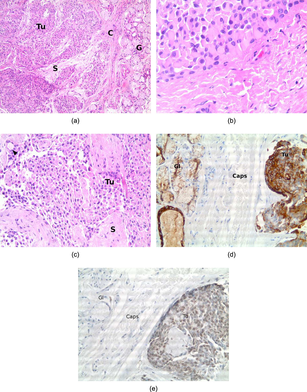
A: Histological view displays a thinly encapsulated tumor (H&E staining, 100×). B: Histological view. Microscopically, homogeneous plasmacytoïd epithelial clusters (H&E staining 400×). C: Histological view showing a rare salivary duct (black arrow) within the tumoral mass. D: Immunostaining positive for cytokeratin AE1–AE3 (healthy gland on the left, ×200). E: Immunostaining positive for PS-100 (healthy gland on the left, ×400). Tu: tumor; S: Stroma; G: healthy Gland; C or Caps: Capsule.
Current usage metrics show cumulative count of Article Views (full-text article views including HTML views, PDF and ePub downloads, according to the available data) and Abstracts Views on Vision4Press platform.
Data correspond to usage on the plateform after 2015. The current usage metrics is available 48-96 hours after online publication and is updated daily on week days.
Initial download of the metrics may take a while.