| Issue |
J Oral Med Oral Surg
Volume 24, Number 2, June 2018
|
|
|---|---|---|
| Page(s) | 76 - 80 | |
| Section | Cas clinique / Short case report | |
| DOI | https://doi.org/10.1051/mbcb/2017031 | |
| Published online | 29 June 2018 | |
Short Case Report
Oral manifestations in Ellis-van Creveld syndrome: a case report
1
Department of Odonto-Stomatology and Maxillofacial surgery, CHU Yalgado Ouedraogo,
Ouagadougou, Burkina Faso
2
Camp General Abd. Sangoulé Latheef Medical Center,
Ouagadougou, Burkina Faso
3
CHU Stomatology Center–Lomé Campus,
Lomé, Togo
4
Muraz de Bobo Dioulasso Center,
Bobo Dioulasso, Burkina Faso
* Correspondence: guiguimdew@yahoo.fr
Received:
1
September
2017
Accepted:
6
December
2017
Introduction: Ellis-van Creveld (EVC) syndrome is an uncommon genetic disease that can be diagnosed at any age. Observation: A case of EVC syndrome was reported in a young 3-year-old female patient presenting chondroectodermal dysplasia, polydactyly, congenital heart defects, damage to the oral mucosa and numerous dental alterations (number, form and structure). Oral management consists of teaching oral hygiene and the prophylactic filling of dental cracks. Discussion: EVC is an autosomal recessive disease. Its diagnosis is only based on clinical features and genetic studies. Conclusion: Dentists should be aware of this syndrome to avoid a late diagnosis and to facilitate a multidisciplinary management.
Key words: Ellis-van Creveld syndrome / genetic / polydactyly
© The authors, 2018
 This is an Open Access article distributed under the terms of the Creative Commons Attribution License (http://creativecommons.org/licenses/by/4.0), which permits unrestricted use, distribution, and reproduction in any medium, provided the original work is properly cited.
Introduction
Ellis-van Creveld (EVC) syndrome or dysplasia chondroectodermal dysplasia, which can be found in Online Mendelian Inheritance in Man entry number 225500, was described in 1940 by Richard W. B Ellis and Simon van Creveld as an autosomal recessive disorder caused by a genetic anomaly located on chromosome 4p16 [1–3]. The prevalence of EVC at birth was estimated to be seven in every 1 000 000 people, and more than 300 cases have been reported in the literature. EVC syndrome occurs mainly in the Amish community in Pennsylvania, USA, with an incidence of five in 1000. It can affect all races, and there is no predilection for sex; however, history of consanguineous marriage is present in 30% cases. The intelligence of affected patients is usually normal [4–6]. Nearly half of these patients die during childhood as a result of cardiopulmonary malformations. For this reason, the life expectancy of patients with EVC is determined by the severity of their congenital heart disease. EVC can be diagnosed by ultrasound from the 18th week of pregnancy onward in the prenatal period and by clinical examination (by a tetrad of symptoms: chondroectodermal dysplasia, polydactyly, cardiac malformations, congenital dental hypoplasia) in the postnatal period [7,8]. Ectodermal dysplasia affects the bones and causes a severe skeletal ossification deficiency, resulting in the shortening of the ribs and limbs and causing pathological fractures [9,10]. The patient often presents with a narrow chest, with an excavation known as a funnel chest, lumbar lordosis, valgum genu, and thinning hair [11]. Polydactyly affects the hands and feet, changing their shape and the number of digits. Congenital cardiac malformations are described in 50–60% patients affected by this syndrome, and they affect mitral valve, and tricuspid valve, and the atrial and ventricular septa and are responsible for the decrease in life expectancy in these patients. Therefore, any dental treatment that carries the risk of infectious endocarditis such as dental extractions, drainage of abscess or perimaxillary cellulitis, frenectomies, scaling, root canal treatments, should be performed under antibiotic prophylaxis to take care of the possible bacteremia resulting from the treatment. This antibiotic prophylaxis is administered in children according to the following protocol: a single dose 30–60 min preoperatively, amoxicillin at 50 mg per kg through intraosseous (IO) infusion or intravenous (IV); and if the patient is allergic to penicillin, administer clindamycin at 20 mg per kg IO or IV. These cardiac malformations are an aggravating factor for repeated bronchopulmonary infections that may result in an EVC patient experiencing respiratory failure, which can result in sudden death [3]. The oral manifestations are diverse and affect not only the soft tissues but also the number, shape, and structure of the teeth. The most frequent manifestation is represented by the labiogingival adhesions, which result in an absence of the labial buccal vestibule [5,11–13]. The anterior part of the lower alveolar ridge is often serrated, and the presence of multiple labial frenulae is noted. The teeth tend to be small and tapered. Molars may exhibit abnormal or additional cusps and sometimes enamel hypoplasia. Congenital oligodontia in temporary and permanent teeth, the presence of supernumerary teeth, dysmorphic natal and neonatal roots, and late dental eruptions have also been reported [11–14]. Dental care depends on each case and requires a multidisciplinary medical team of geneticists, speech therapists, orthopedic surgeons, cardiologists, surgical doctors, pediatricians, and dental specialists. Here we discuss a case of EVC with characteristic and unusual oral and dental manifestations, which will help oral health practitioners to diagnose the syndrome, and refer the patient to the appropriate specialists in time to prevent cardiopulmonary and skeletal complications.
Clinical observation
A 3-year-old child, diagnosed with EVC syndrome, was referred by the pediatrician for dental examination. She was the second child of nonconsanguineous parents and had no congenital anomalies. Her elder brother had no congenital anomalies. She was born by vaginal delivery after an uneventful pregnancy. She had a birth weight of approximately 3200 g, thinning and fine hair, hexadactyly of the hands, and a narrow and disproportionally small thorax (Figs. 1–3). Chondroectodermal syndrome was suspected 7 months after birth. History taking revealed that the patient started walking at the age of 2 years and that her teeth had erupted at the same age. No existence of natal or neonatal teeth has been mentioned. Her medical history revealed that she had repetitive seizures caused by bronchopneumonia, congenital cardiac malformations affecting the mitral and tricuspid valves with a single atrium, corresponding to agenesis of the atrial septum, and a severe insufficiency of skeletal ossification with pathological fractures (Figs. 4–6). On physical examination, the patient was small in stature (77 cm) and weighed 10 kg, had short limbs, had bilateral polydactyly of the hands with hypoplasia of the nails, and had flat feet without polydactyly. Genu valgum was observed. The lumbar spine was normal, without lordosis or kyphosis. In addition, all biochemical, blood, and urinary tests were normal. She had a normal intelligence quotient. The extraoral examination revealed hypertelorism and a marked depression in the middle of the labial philtrum corresponding to maxillary labiogingival fusion. Intraoral examination and care were difficult because the patient did not cooperate well. She was fearful of medical personnel after her multiple hospitalizations. Nevertheless, we noted incomplete dentition (missing teeth 85, 82, 81, 71, 75, 55, 52, 61, 62, and 65) without tooth decay, delayed dental eruption, enamel hypoplasia, conical incisors, and diastemas. Multiple hypertrophic labiogingival frenulae and flanges were visible in the anterior maxillary and mandibular region, rendering the buccal vestibule between the lips and the gingiva nonexistent. (Figs. 7 and 8). The X-ray examination showed dental anomalies such as oligodontia (agenesis of upper lateral incisors and lower central incisors) (Fig. 9). Preliminary dental care consisted of awareness and education on oral hygiene. A treatment for the sealing of the Fissure sealants was performed to prevent decay.
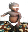 |
Fig. 1 General features of the child. |
 |
Fig. 2 Hands polydactyly and dystrophic nails. |
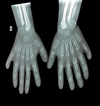 |
Fig. 3 X-ray of the hands showing postaxial polydactyly. |
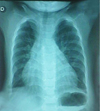 |
Fig. 4 X-ray of the thorax showing narrow chest. |
 |
Fig. 5 Echocardiography showing congenital defects. |
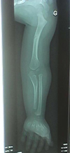 |
Fig. 6 X-ray of the left arm showing pathological fracture of the humerus. |
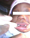 |
Fig. 7 Edentulous mandibular incisor region and oligodontia. |
 |
Fig. 8 Edentulous maxillary incisor region and oligodontia. |
 |
Fig. 9 X-ray facial reconstruction showing oligodontia and conical incisors. |
Discussion
Our patient showed several clinical signs that characterize EVC: bilateral polydactyly affecting the hands; ectodermal dysplasia affecting hair, nails, and teeth; genu valgum; disproportionately small size; and shortened ribs. Other anomalies that may be present include musculoskeletal anomalies, i.e., low shoulders, a narrow rib cage, which frequently causes respiratory distress, similar to our case. Although congenital cardiac malformations occur in only 50–60% cases [1,6,8,9], they have been observed in this case. Mitral and tricuspid valve anomalies and the obvious malformations of the atrial and ventricular septa are some of the malformations described as the main cause of the lower life expectancy in these patients [6]. In our case, the patient had all the characteristics of the symptom tetrad. The fusion of the central part of the upper lip to the maxillary gingival ridge, eliminating the maxillary labial vestibule, and the presence of numerous frenulae attaching the upper lip to the gingiva are characteristics of EVC [5,11–13], which were identified in our case. Patients with EVC may have many tooth anomalies [11–14]. Our patient had several dental anomalies in terms of number (oligodontia), size (microdontia of the incisors), and shape (conical incisors). EVC syndrome requires a multidisciplinary therapeutic approach, i.e., orthopedic corrections, surgical repair of cardiac malformations, and dental intervention for the multiple dental anomalies [4,8,11,15]. Dental treatment includes oral hygiene advice for patients and their parents, and it should include dietary counseling, plaque control methods, periodic consultations, and prophylactic care. Patients with EVC should benefit from dental treatment in early childhood [4]. In this case, treatment included oral hygiene education and prophylactic sealing of the furrow. In this case, treatment included oral hygiene education and fissure sealants [5,11]. These patients may also require surgical and orthodontic treatment, and dental implants may be considered in adulthood [1,11].
Conclusion
EVC syndrome is a rare autosomal recessive disease, which can be diagnosed from its clinical features such as postaxial polydactyly of the hands, dwarfism, fingernails, and dysplastic teeth. A third of these patients die from cardiorespiratory complications in early childhood. Several cases have been reported worldwide, but to our knowledge, this is the first case of EVC with classic symptoms reported in Burkina Faso. The diagnosis and management of EVC syndrome is a great challenge for oral health personnel. Because of genetic and environmental factors altering phenotypes, oral health professionals may see unusual oral and dental anatomy. Therefore, they should update their knowledge to minimize diagnostic errors. To achieve satisfactory functional and aesthetic results, multidisciplinary care planning is necessary for preventing and providing conservative or surgical care to ensure oral health, and to refer patients to the appropriate specialists to prevent cardiac, respiratory, and skeletal complications.
Conflicts of interests
The authors declare that they have no conflicts of interest in relation to this article.
References
- Winter GB, Geddes M. Oral manifestations of chondroectodermal dysplasia (Ellis-van Creveld syndrome). Report of a case. Br Dent J 1967;122:103–107. [PubMed] [Google Scholar]
- Mishra T, Routray SN, Das B. Late survival in Ellis-van Creveld syndrome. A case report. Indian Heart J 2012;64:408–411. [CrossRef] [PubMed] [Google Scholar]
- Allouche M, Ben Ahmed H, Shimi M, Ben Khlil M, Hamdoun M, Baccar H. Syndrome d'Ellis-van Creveld révélé par une mort subite. Cardiologie Tunisienne 2012;08:263–265. [Google Scholar]
- Kamal R, Dahiya P, Kaur S, Bhardwaj R, Chaudhary K. Ellis-van Creveld syndrome: A rare clinical entity. J Oral Maxillofac Pathol 2013;17:132–135. [CrossRef] [PubMed] [Google Scholar]
- Cahuana A, Palma C, Gonzáles W, Geán E. Oral Manifestations in Ellis-van Creveld Syndrome: Report of Five Cases. Pediatr Dent 2004;26:277–282. [PubMed] [Google Scholar]
- Sasalawad SS, Hugar SM, Poonacha KS, Mallikarjuna R. Ellis-van Creveld syndrome. BMJ Case Rep 2013. DOI: 10.1136/bcr-2013-009463 [Google Scholar]
- Ruiz-Perez VL, Goodship JA. Ellis-van Creveld syndrome and Weyers acrodental dysostosis are caused by cilia-mediated diminished response to hedgehog ligands. Am J Med Genet C Semin Med Genet 2009;151C:341–351. [CrossRef] [PubMed] [Google Scholar]
- Baujat G, Le Merrer M. Ellis-van Creveld syndrome. Orphanet J Rare Dis 2007;2:27. [CrossRef] [PubMed] [Google Scholar]
- Shilpy S, Nikhil M, Samir D. Ellis van Creveld syndrome. J Indian Soc Pedod Prevent Dent 2007;25:5–7. [CrossRef] [Google Scholar]
- Alves-Pereira D, Berini-Aytés L, Gay-Escoda C. Ellis-van Creveld syndrome. Case report and literature review. Med Oral Patol Oral Cir Bucal 2009;14:E340–E343. [Google Scholar]
- Souza RC, Martins RB, Okida Y, Giovani EM. Ellis-van Creveld syndrome: oral manifestations and treatment. J Health Sci Inst 2010;28:241–243. [Google Scholar]
- Kalaskar R, Kalaskar AR. Oral manifestations of Ellis-van Creveld syndrome. Contemp Clin Dent 2012;3:S55–S59. [CrossRef] [PubMed] [Google Scholar]
- Ujwala SS, Shibani S, Meghana HC. Ellis-van Creveld syndrome. J Dent 2014;4:641–642. [Google Scholar]
- Shawky RM, Sadik DI, Seifeldin NS. Ellis-van Creveld syndrome with facial dysmorphic features in an Egyptian child. Egypt J Med Human Genet 2010;11:181–185. [CrossRef] [Google Scholar]
- Fukuda A, Kato K, Hasegawa M, Nishimura A, Sudo A, Uchida A. Recurrent knee valgus deformity in Ellis-van Creveld syndrome. J Pediatr Orthop B 2012;21:352–355. [CrossRef] [PubMed] [Google Scholar]
All Figures
 |
Fig. 1 General features of the child. |
| In the text | |
 |
Fig. 2 Hands polydactyly and dystrophic nails. |
| In the text | |
 |
Fig. 3 X-ray of the hands showing postaxial polydactyly. |
| In the text | |
 |
Fig. 4 X-ray of the thorax showing narrow chest. |
| In the text | |
 |
Fig. 5 Echocardiography showing congenital defects. |
| In the text | |
 |
Fig. 6 X-ray of the left arm showing pathological fracture of the humerus. |
| In the text | |
 |
Fig. 7 Edentulous mandibular incisor region and oligodontia. |
| In the text | |
 |
Fig. 8 Edentulous maxillary incisor region and oligodontia. |
| In the text | |
 |
Fig. 9 X-ray facial reconstruction showing oligodontia and conical incisors. |
| In the text | |
Current usage metrics show cumulative count of Article Views (full-text article views including HTML views, PDF and ePub downloads, according to the available data) and Abstracts Views on Vision4Press platform.
Data correspond to usage on the plateform after 2015. The current usage metrics is available 48-96 hours after online publication and is updated daily on week days.
Initial download of the metrics may take a while.


