| Issue |
J Oral Med Oral Surg
Volume 30, Number 1, 2024
|
|
|---|---|---|
| Article Number | 4 | |
| Number of page(s) | 7 | |
| DOI | https://doi.org/10.1051/mbcb/2024007 | |
| Published online | 22 March 2024 | |
Educational Article
Odontogenic orbital cellulitis: literature review
Department of Oral Surgery, Brest University Hospital, Brest, France
* Correspondence: This email address is being protected from spambots. You need JavaScript enabled to view it.
Received:
28
March
2023
Accepted:
25
February
2024
Abstract
Introduction: Odontogenic origin is a rare cause (1.3–5%) of cases of orbital cellulitis, but it can lead to very important morbidity such as blindness or thrombosis of the cavernous sinus. It is therefore important to know how to recognize it. Methods: A literature review was performed. The parameters analyzed included age, sex, ethnicity, clinical presentation, imaging to determine orbital involvement, etiology, microbiology, treatment (medical and/or surgical), and final outcome of each case. Chandler's classification was used to classify the different types of cellulitis. Results: Thirty-five cases of odontogenic orbital cellulitis have been described in literature from 1980 to 2022. In 42.9% of cases, the cellulitis corresponded to an intra-orbital abscess (Chandler stage IV). Thrombosis of the cavernous sinus (stage V) was detected in 5.7% of cases. Periorbital edema (100%), ocular or facial pain (82.9%) and limitation of eye movements (82.9%) were the three most common ophthalmological signs. The anamnesis revealed an element pointing to a dental origin in 97.1% of the cases, the two most frequent being a dental avulsion (20%) or an endodontic treatment (14.3%), in days or weeks preceding the onset of symptoms. Imaging was performed on admission in 94.3% of cases. Regarding the most frequently encountered germs, commensal streptococcus of the oral cavity or anaerobic bacteria were found in 25.7% of cases, and coagulase-negative staphylococcus in 22.9% of cases. In 94.3% of cases, broad-spectrum intravenous antibiotic therapy was initiated as soon as the diagnosis was made. The common feature was the use of metronidazole in 51.4% of cases, combined with a third-generation cephalosporin (11.4%) or amoxicillin-clavulanic acid (8.6%). Orbital drainage was necessary in 71.4% of cases to allow resolution of symptoms, associated with drainage of the maxillary sinus in 45.7% of cases. Finally, the treatment allowed a recovery without sequelae in 80% of cases. Discussion: In case of suspected orbital cellulitis, imaging is crucial to confirm the diagnosis, the type of cellulitis and plan the appropriate surgical treatment. The first step of treatment will be the quick start of a broad spectrum intravenous antibiotic therapy, targeting aerobic and anaerobic bacteria. However, it seems imperative to associate a surgical treatment consisting in a first step of an oral drainage and an elimination of the oral infectious source, as well as an orbital drainage whose approach will have been determined by the imaging. Conclusion: Orbital cellulitis is a rare complication of oral cavity infections, but it must be recognized and treated in time to avoid serious morbidity. An early medical and surgical treatment will usually allow good results and a healing process without sequelae.
Key words: Odontogenic orbital abscess / odontogenic orbital cellulitis
© The authors, 2024
 This is an Open Access article distributed under the terms of the Creative Commons Attribution License (https://creativecommons.org/licenses/by/4.0), which permits unrestricted use, distribution, and reproduction in any medium, provided the original work is properly cited.
Introduction
Orbital cellulitis develops in 70–80% of cases from infection of paranasal sinuses, mainly from the maxillary sinus and ethmoidal sinus [1,2]. Remaining etiologies include periocular trauma, history of surgery, skin infections, dacryocystitis, oral infections and upper respiratory infections [1–4]. Odontogenic origin thus represents only 1.3–5% of cases of orbital cellulitis [2,5–8]. There are three main routes of spread of oral infection to the orbit (Fig. 1). The first route is the most common and corresponds to the spread of oral infections to the maxillary sinus and then to the orbit either by bony erosion of the orbital floor, or via the ethmoidal sinus, or via the infra-orbital canal. In the second route, the infection spreads to the infra-temporal fossa and then reaches the orbital cavity via the inferior orbital fissure. The third route is hematogenous and mainly involves the facial vein and the superior and inferior ophthalmic veins. Indeed, the venous system of the face includes a large number of anastomoses, and the veins lack valves. These two factors together favor the propagation of infections in the form of septic emboli, which can lead to secondary thrombophlebitis, such as cavernous sinus thrombosis [1,9–12].
Orbital cellulitis can cause significant morbidity such as blindness or cavernous sinus thrombosis; therefore, it is essential to know how to recognize it in time in order to be able to perform the appropriate treatment. The purpose of this article was to review the literature on cases of odontogenic orbital cellulitis and to suggest a standardized management of orbital cellulitis of oral origin, based on main medical and surgical approaches reported in the literature to date.
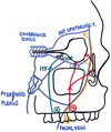 |
Fig. 1 Pathways of propagation of odontogenic orbital cellulitis. ITF: infratemporal fossae; IOF: inferior orbital fissure; IOV: inferior ophthalmic vein. |
Methods
A literature review was conducted, using the following search method:
Time period: 1980 to 2022.
Database searched: PubMed.
Type of publications selected: case reports.
Used keywords were: “odontogenic orbital abscess“, ”odontogenic orbital cellulitis“.
On PudMed with keywords “odontogenic orbital abscess“: 77 results.
On PudMed with keywords ”odontogenic orbital cellulitis“: 65 results.
The titles and abstracts of the studies selected in this preliminary analysis were then listed, and the relevance of each study was determined. Duplicate studies were identified and rejected. The remaining articles were subjected to stricter inclusion and exclusion criteria.
The full texts of the remaining articles were obtained and reviewed on the basis of the following inclusion criteria:
Publications in French and English.
Publications corresponding to case reports of odontogenic orbital cellulitis.
Exclusion criteria included the following:
Case reports dealing with non-odontogenic orbital cellulitis.
Case reports dealing with pathologies other than orbital cellulitis (non-orbital cellulitis of the face, optic neuritis, etc.).
Literature reviews and meta-analyses not including case reports.
Animal studies.
The flow chart shown in Figure 2 represents the systematic review process according to the Preferred Reporting Items for Systematic Reviews and Meta-analyses (PRISMA) guidelines. The different types of cellulitis were classified using the Chandler's classification (Fig. 3), which is based on the extension of the inflammation in relation to the anatomical barriers of the orbit, which are the orbital septum and the periosteum [13].
There are five stages: stage I corresponds to a pre-septal cellulitis, located in front of the orbital septum. Stage II corresponds to a retro-septal cellulitis, also called diffuse orbital cellulitis, which will reach the orbital area behind the septum. Stage III corresponds to a subperiosteal abscess, located between the bony orbital wall and the septum. Stage IV corresponds to an intra-orbital abscess. Stage V corresponds to a thrombosis of the cavernous sinus [13].
Parameters analyzed in the reviewed literature included age, sex, clinical presentation, the realization of an imaging to determine orbital involvement according to the Chandler's classification, etiology, microbiology, treatment, and outcome of each case.
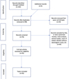 |
Fig. 2 Flow chart showing the methodology used, according to PRISMA guidelines. |
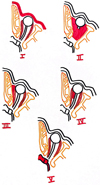 |
Fig. 3 Chandler's classification. |
Results
Thirty-five cases of odontogenic orbital cellulitis have been described in the literature from 1980 to 2022 [1,3–12,14–37]. Among them, the majority were men (65.7%). The mean age at diagnosis was 32.9 (± 19.2) years.
Clinical presentation
Concerning the clinical presentation, and more specifically ophthalmological symptoms, the three most frequently symptoms found in the literature were periorbital edema (100%), ocular or facial pain (82.9%) and limitation of ocular movements (82.9%). The next most frequent findings were peri-ocular erythema (80%), exophthalmos (74.3%), chemosis (65.7%), decreased visual acuity (65.7%), ophthalmoplegia (34.3%), diplopia (31.4%) and relative afferent pupillary deficit (14.3%).
It can also be noted that in two cases (5.7%), a syndrome of the orbital compartment was found, corresponding to a major intraocular hypertonia. In these two cases, the intraocular pressure was higher than 40 mmHg (for a norm of 9–21 mmHg). These cases corresponded both to retro-orbital cellulitis (Chandler type II) [14,24].
Regarding general symptoms, fever was present in a little more than half of the cases (51.4% of cases) and a biological inflammatory syndrome was noted in 45.7% of the cases.
Etiology
In the anamnesis, 97.1% of patients presented a recent oral history or symptoms compatible with an oral inflammatory process (Fig. 4). The two most frequently found were dental avulsion (20% of cases) or endodontic treatment (14.3% of cases), in the days or weeks preceding the onset of symptoms.
Other found elements corresponded to a recent carious removal (5.7%), the recent loss of a dental restoration (2.9%), and a recent root fracture (2.9%). In one case (2.9%), the orbital cellulitis was caused by two associated factors: a fracture of the left zygomatic bone which, after 24 h, led to the decompensation of a chronic endo-periodontal lesion present on the left maxillary first molar, into a retroorbital cellulitis [36]. In another case (2.9%), orbital cellulitis resulted from the infection of a neonatal tooth (a rare phenomenon, defined as a tooth erupting within 30 days of birth) in a neonate [5].
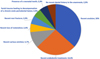 |
Fig. 4 Elements found at the time of anamnesis leading to an oral etiology. |
Imaging
Imaging was performed in 91.4% of cases on admission. In the majority of cases, it was a CT scan of the facial mass (88.6%). A cervico-facial MRI was performed in two cases (5.7%). In two other cases (5.7%), a CT scan of the facial mass was performed after several days, because of an unfavorable evolution under probabilistic antibiotic therapy. Finally, no imaging was performed in one case (2.8%).
Imaging allows the exact type of cellulitis to be defined according to Chandler's classification. The two most frequent types found in the literature corresponded to an intra-orbital abscess (Chandler stage IV) in 42.9% of cases and a sub-periosteal abscess (Chandler stage III) in 25.7% of cases. Retro-septal and pre-septal cellulitis were reported in 20% and 5.7% of cases respectively. Finally, a thrombosis of the cavernous sinus has been demonstrated in two cases (5.7%) [23,31].
Imaging revealed opacification of the maxillary sinus in 20 cases (57.1%), ethmoidal opacification in 12 cases (34.4%) and frontal sinus opacification in 4 cases (11.4%). This sinus opacification is a common CT sign in odontogenic etiology. Periapical lesion is also a common sign, which was found on imaging in 19 cases (54.3%).
Microbiology
The literature reports information on the microbiological results of orbital or sinus pus samples in 28 cases (80%). The sample was positive in 65.7% of cases: a single germ was detected in 42.9% of cases, whereas several germs were found in 22.8% of cases (Fig. 5).
Regarding the most frequently found germs, commensal streptococcus of the oral cavity or anaerobic bacteria were found in 25.7% of cases, and coagulase-negative staphylococcus in 22.9% of cases. In 14.3% of cases, the sample remained negative. This may be related to the use of broad-spectrum antibiotic therapy at diagnosis and prior to sampling.
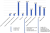 |
Fig. 5 Microbiology. |
Medical and/or surgical management
Broad-spectrum intravenous antibiotic therapy was initiated at diagnosis in 94.3% of cases. Many different combinations have been reported in the literature. The common feature was the use of metronidazole in 51.4% of cases, combined with a third-generation cephalosporin (11.4%) or amoxicillin-clavulanic acid (8.6%). Vancomycin was also used in combination in 17.1% of cases.
Systemic corticosteroid therapy was used in 22.3% of cases. The use of a hypotensive eye-drop was also necessary in 2.9% of cases. Nasal decongestant was used in 8.6% of cases.
Regarding surgical management, orbital drainage was necessary in 71.4% of cases to achieve clinical improvement in combination with antibiotic therapy. Three out of 9 patients (33.3%) required drainage in stages I and II, while this concerned 22 out of 24 patients (91.6%) in stages III and IV, corresponding to the collected stages. The surgical approach was determined by the location of the abscess on imaging. Drainage of the maxillary sinus was associated in 45.7% of cases, by Caldwell Luc approach in half of cases. Then, dental avulsion of the causal tooth was performed in 65.7% of cases. Finally, lateral canthotomy and cantholysis for decompressive purpose were needed in the two cases of cellulitis complicated by an orbital compartment syndrome [14,24]. A transconjunctival retrocaruncular anterior orbitotomy and an endoscopic endonasal decompression of the orbit (by the medial and inferior walls of the orbit) was even necessary in one case, in front of a refractory major intraocular hypertonia [14].
Evolution
The literature reports a complete resolution of symptoms in 80% of cases. On the other hand, in 20% of cases, persistent after-effects are reported: a decrease in residual visual acuity in 14.2% of the cases, and the persistence of diplopia, or headaches and photophobia in 2.9% of the cases.
Discussion
According to literature, the three most frequent signs of odontogenic orbital cellulitis are periorbital edema, ocular or facial pain, and limitation of ocular movements. Visual acuity is normal in 34.3% of cases. These signs are not specific to oral etiology, which is why it is important to look for a dental procedure (avulsion or endodontic treatment) in the days or weeks preceding the onset of symptoms, or for oral symptoms that have recently occurred. It is then essential to perform an imaging of the facial mass when orbital cellulitis is suspected, in order to confirm the diagnosis and the etiology, and to establish the type of cellulitis according to the Chandler's classification. In the majority of reported cases, a CT scan of the facial mass was performed, an examination with good sensitivity that is more accessible than MRI in emergency practice.
Then, the treatment is mainly elaborated in two parts. First, medical treatment, with the quick initiation of broad-spectrum intravenous antibiotic therapy. The use of metronidazole associated with a third-generation cephalosporin has been the most frequently reported combination in the literature, although many other combinations have also been found. It targets both aerobic and anaerobic organisms, the latter being frequently involved in odontogenic orbital cellulitis.
The use of systematic corticosteroid therapy has been reported in 22.3% of cases. The role of this therapy in acute sinusitis and orbital cellulitis was evaluated by Neelam Pushker [38], who compared the use of intravenous antibiotic therapy alone with intravenous antibiotic therapy combined with systemic corticosteroids in patients with orbital cellulitis. The use of high-dose intravenous corticosteroids resulted in faster resolution of symptoms such as fever, pain, periorbital edema, exophthalmos, and limitation of eye movement. In addition, they allowed a faster recovery of visual acuity, but there was no difference in the recovery of long-term visual acuity. Finally, the duration of hospitalization was significantly shorter in the group that received corticosteroid therapy.
According to the literature, surgical treatment associated with this medical treatment is most of the time indispensable. It consists of orbital drainage, which will be almost systematic with most often a drainage of the maxillary sinus in cellulitis associated with Chandler stages III and IV. The approach will be guided by the imaging performed beforehand. Then, it is obviously necessary to treat the cause, and thus to eliminate the oral infectious source (most frequently consisting in the avulsion of the causing tooth) and to perform an oral drainage if it is indicated.
Finally, concerning the evolution, it is interesting to note that among the five cases where there was a sequelae of decreased visual acuity, four cases corresponded to intra-orbital abscesses (type IV). The cause of the persistent visual acuity loss was mostly ischemia of the optic nerve due to compression, or occlusion of the central retinal artery.
Nevertheless, in most cases, appropriate management allows resolution of symptoms without sequelae.
This review of the literature has allowed us to establish a standardized protocol when orbital cellulitis is suspected (Fig. 6).
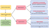 |
Fig. 6 Standard care protocol. |
Conclusion
Orbital cellulitis is a rare complication of infections of oral origin. Nevertheless, it seems essential to know how to recognize it, in order to treat it in time and avoid important after-effects that can go as far as total blindness. An early medical and surgical treatment will usually allow a healing process without sequelae.
Funding
This research did not receive any specific funding.
Conflicts of Interest
The authors declare that they have no conflicts of interest.
Data availability statement
The data that support the findings of this study are available from the corresponding author, Camille GUICHAOUA, upon reasonable request.
Author contribution statement
C. Guichaoua: investigation and writing, S. Genest-Beucher: supervision, S. Boisramé: supervision and revision.
Ethics approval
Ethical approval was not required.
References
- Procacci P, Zangani A, Rossetto A, Rizzini A, Zanette G, Albanese M. Odontogenic orbital abscess: a case report and review of literature. Oral Maxillofac Surg 2017;21:271–279. [CrossRef] [PubMed] [Google Scholar]
- Mbbs MPF, Fraco AAM. Current treatment and outcome in orbital cellulitis: Treatment and outcome in orbital cellulitis. Austr New Zealand J Ophthalmolog 1999;27:375–379. [CrossRef] [PubMed] [Google Scholar]
- de Medeiros EHP, Pepato AO, Sverzut CE, Trivellato AE. Orbital abscess during endodontic treatment: a case report. J Endodontics 2012;38:1541–1543. [CrossRef] [Google Scholar]
- Allan BP, Egbert MA, Myall RWT. Orbital abscess of odontogenic origin. Case report and review of the literature. Int J Oral Maxillofac Surg 1991;20:268–270. [Google Scholar]
- White M. et al. Neonatal tooth infection resulting in subperiosteal orbital abscess: a case report. Int J Pediatric Otorhinolaryngol 2021;140:110524. [CrossRef] [Google Scholar]
- Geusens J, Dubron K, Meeus J, Spaey Y, Politis C. Subperiosteal orbital abscess from odontogenic origin: a case report. Int J Surg Case Rep 2020;73:263–267. [CrossRef] [PubMed] [Google Scholar]
- Eltayeb AS, Karrar MA, Elbeshir EI. Orbital subperiosteal abscess associated with mandibular wisdom tooth infection: a case report. J Maxillofac Oral Surg. 2019;18:30–33. [CrossRef] [PubMed] [Google Scholar]
- Janakarajah N, Sukumaran K. Orbital cellulitis of dental origin: case report and review of the literature. Br J Oral Maxillofac Surg. 1985;23:140–145. [CrossRef] [Google Scholar]
- Muñoz-Guerra MF, González-García R, Capote AL, Escorial V, Gías LN. Subperiosteal abscess of the orbit: an unusual complication of the third molar surgery. Oral Surg Oral Med Oral Pathol Oral Radiol Endodontol 2006;102:e9–e13. [CrossRef] [Google Scholar]
- Poon TL, Lee WY, Ho WS, Pang KY, Wong CK. Odontogenic subperiosteal abscess of orbit: a case report. J Clin Neurosci 2001;8:469–471. [CrossRef] [PubMed] [Google Scholar]
- Thakar M, Thakar A. Odontogenic orbital cellulitis: Report of a case and considerations on route of spread. Acta Ophthalmolog Scand 2009;73:470–471. [Google Scholar]
- Grimes D. Case report: dental infection leading to orbital cellulitis. Dental Update 2006;217–220. [CrossRef] [PubMed] [Google Scholar]
- Chandler JR, Langenbrunner, David J, Stevens ER. The pathogenesis of orbital complications in acute sinusitis. Laryngoscope 1970;80:1414–1428. [CrossRef] [PubMed] [Google Scholar]
- Rothschild MI, Pinheiro-Neto CD, Rubinstein TJ. Odontogenic abscess with orbital extension through the inferior orbital fissure treated with bony decompression. Ophthalmic Plastic Reconstruc Surg 2020;36:e131–e134. [CrossRef] [PubMed] [Google Scholar]
- Tavakoli M, Bagheri A, Faraz M, Salehirad S, Roghaee S. Orbital cellulitis as a complication of mandibular odontogenic infection. Ophthalmic Plastic Reconstruc Surg 2013;29:e5–e7. [CrossRef] [PubMed] [Google Scholar]
- Hughes E, Wynne N, Quinn S, Fulcher T. Odontogenic orbital abscess with intracranial and pulmonary involvement. Orbit 2017;36:459–461. [CrossRef] [PubMed] [Google Scholar]
- Kim I-K., Kim J-R., Jang K-S., Moon Y-S., Park S-W. Orbital abscess from an odontogenic infection. Oral Surg Oral Med Oral Pathol Oral Radiol Endodontol 2007;103:e1–e6. [CrossRef] [Google Scholar]
- Mansour AM, Kheir-Jurdi W, Hadi UE, Awar G. Odontogenic abscess mimicking acute dacryocystitis. BMJ Case Rep 2017 bcr- 2016–218560. [Google Scholar]
- Sakkas N, Schoen R, Schmelzeisen R. Orbital abscess after extraction of a maxillary wisdom tooth. Br J Oral Maxillofac Surg 2007;45:245–246. [CrossRef] [PubMed] [Google Scholar]
- Koch F, Breil P, Marroquin BB, Gawehn J, Kunkel M. Abscess of the orbit arising 48 h after root canal treatment of a maxillary first molar. Int Endod J 2006;39:657–664. [CrossRef] [PubMed] [Google Scholar]
- Stübinger S, Leiggener C, Sader R, Kunz C. Intraorbital abscess. J Am Dental Assoc 2005;136:921–925. [CrossRef] [Google Scholar]
- de Assis-Costa MDM, Santos GS, Maciel J, Sonoda CK, de Melo WM. Odontogenic infection causing orbital cellulitis in a pediatric patient. J Craniofac Surg 2013;24:e526–e529. [CrossRef] [PubMed] [Google Scholar]
- Allegrini D, Reposi S, Nocerino E, Pece A. Odontogenic orbital cellulitis associated with cavernous sinus thrombosis and pulmonary embolism: a case report. J Med Case Rep 2017;11:164. [CrossRef] [PubMed] [Google Scholar]
- Park CH, Jee DH, La TY. A case of odontogenic orbital cellulitis causing blindness by severe tension orbit. J Korean Med Sci 2013;28:340. [CrossRef] [PubMed] [Google Scholar]
- Arunkumar KV. Orbital infection threatening blindness due to carious primary molars: an interesting case report. J Maxillofac Oral Surg 2016;15:72–75. [CrossRef] [PubMed] [Google Scholar]
- Wysluch A, Maurer P, Ast J, Kunkel M. Orbital complications due to an acute odontogenic focus in a child. A case report. Oral Surg Oral Med Oral Pathol Oral Radiol Endodontol 2009;107:e39–e42. [CrossRef] [Google Scholar]
- Rosen D, Ardekian L, Abu El-Naaj I, Fischer D, Peled M,Laufer D. Orbital infection arising from a primary tooth: a case report. Int J Paediatric Dent 2001;10:237–239. [Google Scholar]
- Mitchell CS, Nelson MD. Orofacial abscesses of odontogenic origin in the pediatric patient: report of two cases. Pediatr Radiol 1993;23:432–434. [CrossRef] [PubMed] [Google Scholar]
- Henry H, Hughes V, Larned DC. Odontogenic Infection of the Orbit: Journal of Oral and Maxillofacial Surgery. 1992;50:172–8. [Google Scholar]
- Akhaddar A. et al. Orbital abscess associated with sinusitis from odontogenic origin. Intern Med 2010;49:523–524. [CrossRef] [PubMed] [Google Scholar]
- Ogundiya DA, Keith DA, Mirowski J. Cavernous sinus thrombosis and blindness as complications of an odontogenic infection: report of a case and review of literature. J Oral Maxillofac Surg 1989;47:1317–1321. [CrossRef] [PubMed] [Google Scholar]
- Robbins KT, Tarshis LM. Blindness: A complication of odontogenic sinusitis. Otolaryngol Head Neck Surg 1981;89:938–940. [CrossRef] [PubMed] [Google Scholar]
- Li E, Distefano A, Sohrab M. Necrotizing orbital cellulitis secondary to odontogenic Streptococcus constellatus. Ophthalmic Plastic Reconstruc Surg 2018;34:e160–e162. [CrossRef] [PubMed] [Google Scholar]
- Mehra P, Caiazzo A, Bestgen S. Odontogenic sinusitis causing orbital cellulitis. J Am Dent Assoc 1999;130:1086–1092. [CrossRef] [PubMed] [Google Scholar]
- Yan W, Chakrabarti R, Choong J, Hardy T. Orbital cellulitis of odontogenic origin. Orbit 2015;34:183–185. [CrossRef] [PubMed] [Google Scholar]
- Hovinga J, Christiaans BJ. Odontogenic infection leading to orbital cellulitis as complication of fracture of the zygomatic bone. J Cranio-Maxillofac Surg 1987;15:254–257. [CrossRef] [Google Scholar]
- Houle AN, Pham C, Valikodath N, Elmowitz JS, Callahan N. Odontogenic subperiosteal abscess of the lateral orbit: timely recognition and management. Eur J Dent 2021;15:802–805. [CrossRef] [PubMed] [Google Scholar]
- Pushker N, Tejwani LK, Bajaj MS, Khurana S, Velpandian T, C handra M. Role of oral corticosteroids in orbital cellulitis. Am J Ophthalmol 2013;156: 178–183.e1. [Google Scholar]
All Figures
|
Fig. 1 Pathways of propagation of odontogenic orbital cellulitis. ITF: infratemporal fossae; IOF: inferior orbital fissure; IOV: inferior ophthalmic vein. |
| In the text | |
|
Fig. 2 Flow chart showing the methodology used, according to PRISMA guidelines. |
| In the text | |
|
Fig. 3 Chandler's classification. |
| In the text | |
|
Fig. 4 Elements found at the time of anamnesis leading to an oral etiology. |
| In the text | |
|
Fig. 5 Microbiology. |
| In the text | |
|
Fig. 6 Standard care protocol. |
| In the text | |
Current usage metrics show cumulative count of Article Views (full-text article views including HTML views, PDF and ePub downloads, according to the available data) and Abstracts Views on Vision4Press platform.
Data correspond to usage on the plateform after 2015. The current usage metrics is available 48-96 hours after online publication and is updated daily on week days.
Initial download of the metrics may take a while.