| Issue |
J Oral Med Oral Surg
Volume 25, Number 4, 2019
|
|
|---|---|---|
| Article Number | 34 | |
| Number of page(s) | 4 | |
| Section | Article pédagogique / Educational article | |
| DOI | https://doi.org/10.1051/mbcb/2019015 | |
| Published online | 17 September 2019 | |
Educational Article
Oral and skin manifestations of tuberous sclerosis complex
1
Aix Marseille Univ, APHM, La Timone, Service d'Odontologie, 264 rue Saint Pierre, 13385 Marseille Cedex 5, France
2
UMR 7268 ADES, Aix-Marseille/EFS/CNRS, faculté de médecine-secteur nord, Boulevard Pierre-Dramard, 13344 Marseille Cedex 15, France
3
Centre Massilien de la Face, 24 avenue du Prado, 13006 Marseille, France
4
UMR_S910. Centre de génétique médicale de Marseille, Aix Marseille Univ, Campus Timone, 27 Bd J Moulin, 13385 Marseille Cedex 5, France
* Correspondence: This email address is being protected from spambots. You need JavaScript enabled to view it.
Received:
6
June
2018
Accepted:
29
May
2019
Abstract
Tuberous sclerosis complex is a genetic disease characterized by multisystemic hamartomas with variable and non-specific clinical manifestations. The disease is associated with mutations of genes encoding the proteins hamartin and tuberin. The hamartin/tuberin complex plays an anti-tumor function by inhibiting mammalian target of rapamycin. The diagnostic criteria for the disease were reviewed at a consensus conference in 2012. Evidence of mutations of tuberous sclerosis complex 1 or 2 genes has become a clinical and independent diagnostic criterion. Among the clinical criteria used, two oral criteria include the presence of three or more enamel pits and the presence of two or more oral fibromas. Several dermatological criteria are included within these criteria and are of interest in our specialty when these are localized at the cephalic extremity.
Key words: tuberous sclerosis / oral manifestations / skin manifestations
© The authors, 2019
 This is an Open Access article distributed under the terms of the Creative Commons Attribution License (http://creativecommons.org/licenses/by/4.0), which permits unrestricted use, distribution, and reproduction in any medium, provided the original work is properly cited.
Introduction
Tuberous sclerosis (Bourneville disease) or tuberous sclerosis of the brain is a genetic disorder characterized by multisystemic hamartomas with variable and non-specific clinical manifestations. The diagnostic criteria for the disease were updated in a consensus conference in 2012 [1] The most significant development was the inclusion of a genetic criterion allowing the diagnostician to make diagnosis independently. Among the clinical diagnostic criteria, two oral criteria were included as minor criteria. The presence of three or more enamel pits was introduced as a diagnostic criterion during this revision, and the presence of gingival fibromas was replaced by the presence of two or more oral fibromas. Moreover, several dermatological criteria were included within the major and minor diagnostic criteria, which are of interest in our specialty, particularly when they are localized around the cephalic extremity.
The aim of this article was to emphasize the manifestations and management of oral and dermatological localizations of tuberous sclerosis.
Definition, epidemiology, and genetics
The first description of the disease was made by von Recklinghausen in 1862 [1], and in 1880 Bourneville gave it the name tuberous sclerosis. Its prevalence has been estimated at 1 case per 10,000 to 25,000 individuals [2,3]. This prevalence is probably underestimated due to undiagnosed cases [4]. Tuberous sclerosis belongs to the group of classical phacomatoses [2] with neurofibromatosis types 1 and 2, Sturge–Weber–Krabbe syndrome, Von Hippel–Lindau disease, and various neuroectodermal dysembryoplasias. It is an autosomal dominant genetic disease with almost complete penetrance; however, two-thirds of individuals develop the disease following pathogenic de novo variation [2,3]. The disease is associated with a pathogenic mutation of two genes. However, despite the advances in diagnostic techniques, no mutation is detected in 15%–20% of the cases, not excluding diagnosis [1,4]. In 31% of the patients, a mutation is identified in the tuberous sclerosis complex 1 gene (TSC1) located on chromosome 9 (9q34), and in 69% of the patients, a mutation is identified the TSC2 gene located on chromosome 16 (16p13.3) [4,5]. The genes TSC1 and TSC2 respectively encode hamartin and tuberin [4] that combine to form a hamartin–tuberin complex. Mutation of either of the proteins renders the complex inactive [4]. This complex shows anti-tumor activity by inhibiting the activity of mammalian target rapamycin target (mTOR) protein [4], which is a regulatory kinase for cell proliferation and growth [4].
Diagnosis
Tuberous sclerosis has a predominantly neurocutaneous expression characterized by multisystemic hamartomas associated with neuropsychiatric manifestations such as mental retardation and epilepsy [2]. Many symptoms associated with tuberous sclerosis are not pathognomonic, which poses diagnostic difficulties. The Washington International Consensus Conference in 2012 [1,5] modified the diagnostic criteria for this disease.
A genetic diagnostic criterion was introduced during this revision [1,5]. The presence of pathogenic mutations of the TSC1 or TSC2 genes allows for a definitive diagnosis of tuberous sclerosis, independent of the associated clinical manifestations.
The second diagnostic criterion is clinical [1]. Clinical manifestations are grouped into 11 major and 6 minor criteria (Tab. I). Diagnosis is considered definitive in the presence of two major clinical criteria or one major clinical criterion and two minor clinical criteria. Diagnosis is considered possible in the presence of a major clinical criterion or at least two minor clinical criteria. Lymphangioleiomyomatosis and angiomyolipoma constitute, when concomitant, a single major criterion and not two major criteria. Among the minor clinical criteria, two are oral diagnostic criteria: The presence of at least two intraoral fibromas and at least three enamel pits.
Diagnostic criteria for tuberous sclerosis (Bourneville disease) according to the 2012 consensus conference. Red: oral criteria; blue: dermatological criteria.
Oral manifestations
Oral manifestations of tuberous sclerosis are noted in 11%–56% of the patients [6]. These are sometimes barely perceptible or at other times remarkable. Oral lesions are usually diagnosed between the age of 4 to 10 years or during puberty [6,7].
Enamel pits are the most common oral manifestation of the disease and are present in almost all patients [1,10,14]. These correspond to enamel hypoplasia [6,10] without associated dentinal involvement [6,7]. Their diameter varies from 4 to 100 µm. These are detected clinically or by retroalveolar radiography when radiolucent [12]. These increase the risk of cavities [10]. Treatment is necessary when enamel pits are symptomatic, decayed, or unsightly [5].
Oral fibromas are the second most common manifestation of tuberous sclerosis (Fig. 1). They are localized most often on the maxillary anterior gingiva but can be observed on the cheeks, lips, edge of the lip, tongue, or palate [7–11]. According to the studies, their prevalence is 11%–69% [6,9,10,12–15] and average diameter is 5 mm [6,10]. These fibromas can achieve gingival growth [14], which can be confused with a drug-related etiology when anticonvulsant therapy, particularly phenytoin, is concomitantly prescribed [5,7,10,12,13]. According to Curi et al. [16], the differential diagnosis is based on the purely gingival involvement in cases of increased drug dose without lesions affecting any other mucous membranes contrary to the tuberous sclerosis. Removal of these fibromas is indicated in case of an increase in size or aesthetic or functional discomfort with associated bleeding [5]. This excision can be achieved via surgery, CO2 laser vaporization, or electrocauterization [5,6].
Other oral manifestations have been described in tuberous sclerosis cases. According to Gavren et al. [3], tuberous sclerosis can be associated with cleft lip and palate, high-arched palate, bifid uvula, and macroglossia. Cases of bony desmoid fibroids [6,13,17], odontogenic fibroids [10], and myxomas [10] have been reported, with sporadic cases of oral angiomyolipoma [12]. According to Barron et al. [12], the treatment of intraosseous fibroblastic lesions is performed by curettage and enucleation. Oral monitoring every 6 months is recommended [5,6,10].
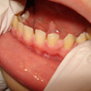 |
Fig. 1 Gingival fibromas of the mandible. |
Dermatological manifestations
Dermatological manifestations are lesions of interest for an oral surgeon. These contribute to the diagnosis of the disease as four manifestations are part of the major clinical criteria (hypomelanotic macules at least 5 mm in diameter (n ≥ 3), facial angiofibromas (n ≥ 3) or fibrous cephalic plaques, nongelial fibroids (n ≥ 2), and shagreen patch) and one is a part of the minor clinical criteria (“confetti-like” skin lesions) [5].
Hypomelanotic macules (Fig. 2) are found in 90% of the patients [5]. These may be present from birth or may appear during childhood [1,5]. These do not generally require treatment unless they affect a patient's appearance. Topical mTOR inhibitors have been successfully used for management [4,6]. Facial angiofibromas (Fig. 3) are reported in more than 70% of the patients. These appear in the form of reddish fibrovascular papules. These angiofibromas usually appear before the age of 10 years and are usually a pathognomonic sign [5]. They are localized around the nose, cheeks, and chin, with a classically symmetrical arrangement. Their numbers increase at puberty [3]. Their management is based on topical mTOR inhibitors, vascular or surgical lasers, and resection [4,6]. A cephalic fibrous plate, usually unilateral, is found in 25% of the patients [5]. Its histology is similar to that of angiofibromas [1]. Surgery is proposed when it increases in size and/or is unsightly [6]. Ungular fibromas are found in 20% of the children, but this percentage can increases over time to become 80% of the adults [1]. Laser or surgical excision is indicated when their size is >3 mm [6]. A “shagreen patch” appears in more than 50% of the patients, usually before the age of 10 years. It is in the form of a large bumpy lesion with a so-called orange peel surface, usually located on the lower back [1]. “Confetti-like” skin lesions occur as millimetric hypomelanotic macules [5]. They differ from hypomelanotic macules in their smaller diameter and greater number [1]. These lesions, quite common in adults, are useful for diagnosis when they are congenital or appear before the age of 10 years [5].
Annual skin monitoring is recommended in children and should be individually adapted according to the clinical course [6].
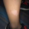 |
Fig. 2 Hypomelanotic macula of the right leg. |
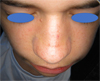 |
Fig. 3 Facial angiofibromas. |
Conclusion
Tuberous sclerosis (Bourneville disease) is a rare pathology with potentially severe consequences. Because of its rarity, oral clinical manifestations are often overlooked by different oral specialists. Although non-specific, two oral signs form part of the clinical diagnostic criteria for the disease, as determined at the last consensus conference (2012). The presence of enamel pits or multiple oral fibromas should evoke the diagnosis of tuberous sclerosis. Oral specialists should therefore adopt a multidisciplinary approach both in terms of diagnosis and management.
Conflicts of interests
The authors declare that they have no conflicts of interest in relation to the publication of this article.
References
- Northrup H, Krueger D. Tuberous Sclerosis Complex Surveillance and Management: Recommendations of the 2012 International Tuberous Sclerosis Complex Consensus Conference. Pediatr Neurol 2013;49:255–265. [Google Scholar]
- Orphanet: Sclérose tubéreuse de Bourneville. Available at: http://www.orpha.net/consor/cgi-bin/OC_Exp.php?Lng=FR&Expert=805. (Accessed: 5th May 2018) [Google Scholar]
- Gavren BA, Lumerman H, Cardo VA, Schmidt BL. Multiple pigmented lesions of the lower lip. J Oral Maxillofac Surg Off J Am Assoc Oral Maxillofac Surg 2002;60:438–445. [CrossRef] [Google Scholar]
- Sasongko TH, Ismail NF, Zabidi-Hussin Z. Rapamycin and rapalogs for tuberous sclerosis complex. Cochrane Database Syst Rev 2016;7:CD011272. [PubMed] [Google Scholar]
- Teng JM, Cowen EW, Wataya-Kaneda M, Gosnell ES, Witman PM, Hebert AA, et al. Dermatologic and Dental Aspects of the 2012 International Tuberous Sclerosis Complex Consensus Statements. JAMA Dermatol 2014;150:1095–1101. [CrossRef] [PubMed] [Google Scholar]
- Nico MM, Hammerschmidt M, Lourenço SV. Oral mucosal manifestations in some genodermatoses: correlation with cutaneous lesions. Eur J Dermatol 2013;23:581–591. [Google Scholar]
- López E, Escovich L, Vigna A. Tuberous sclerosis: presentation of a clinical case with oral manifestations. Med Oral 2003; 8:122–128. [PubMed] [Google Scholar]
- Araújo Lde J, Muniz GB, Santos E, Ladeia JP, Martelli H Jr, Bonan PR. Tuberous sclerosis complex diagnosed from oral lesions. Sao Paulo Med J 2013;131:351–355. [CrossRef] [PubMed] [Google Scholar]
- Damm DD, Tomich CE, White DK, Drummond JF. Intraosseous fibrous lesions of the jawsA manifestation of tuberous sclerosis. Oral Surg Oral Med Oral Pathol Oral Radiol Endod 1999;87:334–340. [CrossRef] [PubMed] [Google Scholar]
- Harutunian K, Figueiredo R, Gay-Escoda C. Tuberous sclerosis complex with oral manifestations: a case report and literature review. Med Oral Pathol Oral Cir Bucal 2011;16:e478–e481. [CrossRef] [Google Scholar]
- Scully C. Oral mucosal lesions in association with epilepsy and cutaneous lesions: the Pringle-Bourneville syndrome. Int J Oral Surg 1981;10:68–72. [CrossRef] [PubMed] [Google Scholar]
- Barron RP, Kainulainen VT, Forrest CR, Krafchik B, Mock D, Sandor GK. Tuberous sclerosis: clinicopathologic features and review of the literature. J Craniomaxillofac Surg 2002;30:361–366. [CrossRef] [PubMed] [Google Scholar]
- López-López J, Rodriguez-de-Rivera-Campilo E, Marques-Soares MS, Finestres-Zubeldia F, Chimenos-Küstner E, Rosello-Llabres X. Tuberous sclerosis and its oral manifestations. A clinical case. Med Oral 2004;9:216–223. [PubMed] [Google Scholar]
- Sparling JD, Hong CH, Brahim JS, Moss J, Darling TN. Oral findings in 58 adults with tuberous sclerosis complex. J Am Acad Dermatol 2007;56:786–790. [CrossRef] [PubMed] [Google Scholar]
- Lygidakis NA, Lindenbaum RH. Oral fibromatosis in tuberous sclerosis. Oral Surg Oral Med Oral Pathol 1989;68:725–728. [Google Scholar]
- Curi MM, Cardoso CL, Ikuta CRS, Koga DH, Zardetto C. Tuberous Sclerosis: A case report with oral manifestation. Int J Odontostomatol 2014;8:185–189. [CrossRef] [Google Scholar]
- Kennedy RA, Thavaraj S, Diaz-Cano S. An overview of autosomal dominant tumour syndromes with prominent features in the oral and maxillofacial region. Head Neck Pathol 2017; 11:364–376. [CrossRef] [PubMed] [Google Scholar]
All Tables
Diagnostic criteria for tuberous sclerosis (Bourneville disease) according to the 2012 consensus conference. Red: oral criteria; blue: dermatological criteria.
All Figures
|
Fig. 1 Gingival fibromas of the mandible. |
| In the text | |
|
Fig. 2 Hypomelanotic macula of the right leg. |
| In the text | |
|
Fig. 3 Facial angiofibromas. |
| In the text | |
Current usage metrics show cumulative count of Article Views (full-text article views including HTML views, PDF and ePub downloads, according to the available data) and Abstracts Views on Vision4Press platform.
Data correspond to usage on the plateform after 2015. The current usage metrics is available 48-96 hours after online publication and is updated daily on week days.
Initial download of the metrics may take a while.