| Issue |
J Oral Med Oral Surg
Volume 25, Number 2, 2019
|
|
|---|---|---|
| Article Number | 21 | |
| Number of page(s) | 5 | |
| Section | Cas clinique et revue de la littérature / Up-to date review and case report | |
| DOI | https://doi.org/10.1051/mbcb/2019005 | |
| Published online | 01 May 2019 | |
Up-to Date Review And Case Report
Oral nodular fasciitis – A case report with a diagnostic schema
1
Department of Oral Pathology and Microbiology, Manipal College of Dental Sciences, Manipal, Manipal Academy of Higher Education, Manipal, Karnataka, India
2
Department of Pathology, Kasturba Medical College, Manipal, Manipal Academy of Higher Education, Manipal, Karnataka, India
3
Department of Oral and Maxillofacial Surgery, Manipal College of Dental Sciences, Manipal, Manipal Academy of Higher Education, Manipal, Karnakata, India
4
Department of Oral Pathology and Microbiology, Manipal College of Dental Sciences, Manipal, Manipal Academy of Higher Education, Manipal, Karnataka, India
* Correspondence: This email address is being protected from spambots. You need JavaScript enabled to view it.
Received:
8
December
2018
Accepted:
18
March
2019
Abstract
Introduction: The spectrum of myofibroblastic lesions of the oral cavity ranges from reactive to benign to malignant lesions with overlapping histopathologic and immunohistologic characteristics posing a diagnostic dilemma. Observation: A 30-year-old male presented with a spontaneous swelling over the right lower buccal gingiva giving a clinical suspicion of a benign mesenchymal tumor. The lesion presented with a varied biphasic microscopic appearance that posed as a challenge for diagnosis. Commentaries: The incisional biopsy of the lesion showed a highly collagenous stroma with spindle-shaped cells, while the excision biopsy revealed myxoid and hyalinized stroma. A panel of markers comprising of SMA (smooth muscle actin). CD-34, β-Catenin, and Alcian blue stain was employed to arrive at a diagnosis. Conclusion: Most myofibroblastic lesions present with diverse histological appearance which warrants a thorough assessment of the cellular and stromal components for an accurate diagnosis.
Key words: spindle cells / nodular fasiitis / myofibroblasts / myxoid degeneration / myofibroma
© The authors, 2019
 This is an Open Access article distributed under the terms of the Creative Commons Attribution License (http://creativecommons.org/licenses/by/4.0), which permits unrestricted use, distribution, and reproduction in any medium, provided the original work is properly cited.
Introduction
Nodular fasciitis (NF) is a benign reactive soft tissue lesion that was first described by Konwaler in 1955 as a subcutaneous pseudosarcomatous fibromatosis [1]. NF most commonly present in the extremities, trunk, head and neck region. The incidence of NF in the oral cavity is very rare and it occurs mostly in the buccal mucosa, alveolar mucosa, tongue and upper lip [2–4]. Nodular fasciitis occurs equally among both males and females in the fourth and fifth decades of life. It presents as a rapidly growing mass that can grow at a rate of approximately 4 cms in one month. It is firm and fixed on palpation and the surface of the growth may be ulcerated [3,5].
The pathogenesis of NF is unknown, although trauma followed by inflammation is implicated as a causative factor [6]. Although NF is a benign reactive lesion, due to its rapid growth, high cellularity and high mitotic rate it is often misdiagnosed as a sarcoma [7]. NF has to be differentiated from schwannoma, myofibroma, solitary fibrous tumor, fibrosarcoma, leiomyosarcoma, neurofibroma and malignant fibrous histiocytoma [4,6,8]. The positivity of tumor cells for smooth muscle actin (α-SMA), muscle specific actin (HHF-35) and vimentin has established the myofibroblastic origin of NF [9].
Here, we present a case of NF that occurred on the alveolar mucosa, a rare site for the lesion. The lesion presented with a biphasic histological pattern which posed as a challenge for its diagnosis. The histochemical and immunohistochemical analysis that was carried out to arrive at the diagnosis is described.
Observation
A 30-year-old male patient visited the dental clinic with a complaint of a growth in the right lower teeth region since 3 months, which gradually increased in size. There was no history of pain, pus discharge or mobility of teeth. The medical history was otherwise not remarkable.
On clinical examination, the lesion presented as a lobulated growth measuring approximately 3 × 3 cm in the buccal vestibule in relation to the right mandibular second premolar, first molar and second molar (Fig. 1). The lesion was pink in color, with well-defined margins, was firm in consistency and mildly tender on palpation. There was no evidence of induration or discharge from the lesion. Based on the clinical presentation, a provisional diagnosis of benign mesenchymal tumor such as fibroma, irritational fibroma and peripheral giant cell granuloma was proffered. An incisional biopsy of the soft tissue mass was obtained for histopathological examination.
The hematoxylin and eosin stained sections revealed a highly cellular and collagenous stroma comprising spindle-shaped cells arranged in a storiform pattern (Fig. 2a). The spindle-shaped cells were elongated with an oval nuclei and eosinophilic cytoplasm. The connective tissue stroma comprised of plump fibroblasts, fibrocytes, variable number of blood vessels, and areas of hyalinization. Chronic inflammatory cells were seen in the perivascularly.
On Immunohistochemical analysis, the cytoplasm of the spindle cells expressed SMA (clone IA4, Dako) (Fig. 2b) but did not express CD-34 (clone QBE 10, Dako) (Fig. 2c) or β-Catenin (EP 35, Pathnsitu) (Fig. 2d). The findings were in favor of solitary myofibroma. The entire lesion was subsequently excised.
The hematoxylin and eosin stained tissue section of the operative piece showed immature-appearing myofibroblasts admixed with myxomatous and hyalinized areas. The cells had an oval pale staining nuclei with prominent nucleoli (Figs. 3a and 3b). The intervening matrix was rich in mucopolysacchrides which stained readily with Alcian blue (Fig. 3c). Based on the histochemical and immuhistochemical profile of the lesional tissue, a final diagnosis of nodular fasciitis was given (Fig. 4). The patient is on regular follow-up and there is no sign of recurrence since the last 8 months.
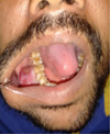 |
Fig. 1 A 3 × 3 cm nodular growth on the right mandibular alveolus. |
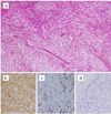 |
Fig. 2 Photomicrograph showing a) A collagenous stroma comprising of numerous proliferating spindle cells arranged in a biphasic pattern (H&E 4X), (b) Spindle cells showing cytoplasmic expression of smooth muscle actin (IHC-10X), c) CD 34 expression present in endothelial cells but absent in spindle cells (IHC-10X), d) Spindle-shaped cells lacking the expression of β-Catenin (IHC-10X). |
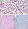 |
Fig. 3 Photomicrograph showing a) Fascicles of proliferating spindle cells in a hyalinized stroma with extravasated RBC's and cleft-like spaces (H&E-20X), b) Hyalinized stroma (H&E 10X) and c) stroma rich in mucopolysaccharide (Alcian blue stain – 10X). |
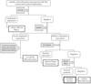 |
Fig. 4 A decision tree depicting the diagnostic approach. |
Commentary
Nodular fasciitis is a benign reactive lesion of the fibrous connective tissue with proliferation of fibroblasts and myofibroblasts arranged in short fascicles [5]. This benign reactive lesion is said to arise from the fascia and lesion extends into the septae of the subcutaneous fat [10,11].
Allen et al. had described four typical histological features of NF, numerous spindle-shaped fibroblasts arranged in whorled fascicles, small clefts separating the fibroblasts, scattered extravasated red blood cells and a mucoid interstitial ground substance [12]. The spindle cells exhibiting mitotic activity, areas of myxoid degeneration, fibrosis and hyalinization, and the presence of giant cells are additional features of NF [13,14]. The myxoid stroma gives rise to a “feathery pattern” in early lesions and with time the lesion gets progressively hyalinised [15]. The histological features that were seen in the present case also included spindle-shaped cells, areas of hyalinization, areas of myxoid degeneration and a mucoid stroma.
Due to the high proliferative capacity of the nodular fasciitis, it has to be differentiated from malignant lesions such as myxofibrosarcoma, low-grade fibromyxoid sarcoma, and myofibroblastic sarcoma. The lack of an invasive growth pattern, absence of nuclear hyperchromatism, cellular pleomorphism and atypical mitotic figures and areas of necrosis helped in diagnosis of a benign lesion [9]. In addition, the expression of SMA is not uniform and is restricted in these sarcomatous lesions [15].
The histological features that differentiate nodular fasciitis from other benign spindle cell lesions is given in Table I. Cytoplasmic CD-34 expression is consistent with solitary fibrous tumor (SFT), however, in the present case the expression was restricted to the capillaries in between the tumor cells [15]. Moreover, a nuclear expression of β-catenin is characteristic of fibromatosis and SFT which was negative in the present case [16].
In this case, the spindle-shaped myofibroblast-like cells stained positive for smooth muscle actin. This was also observed in the cases that were reported by lloyd et al., Ikebe et al. and Souza et al. [4,17,18] however, NF has to be distinguished from other benign tumors of myofibroblastic origin. Inflammatory myofibroblastic tumor (IMT) is generally larger than NF, tends to occur at a younger age, and is composed of longer fascicles of spindle cells in an inflammatory background that is rich in plasma cells. IMT is a tumor that commonly occurs in the lung, however, the presence of this tumor in the mandible has been reported [19].
NF can be distinguished from solitary myofibroma on the basis of its “biphasic zoning” phenomenon. The biphasic zoning phenomenon is seen in solitary myofibroma and refers to the presence of light-staining collageneous, hyalinized areas adjacent to dark-staining hemangiopericytoma-like areas. SMF also lack the mucopolysaccharide rich stroma as demonstrated in the present case thus directing the diagnosis to nodular fasciitis.
Although nodular fasciitis is regarded as a self-limiting lesion, relocation of USP6 gene was detected. This was also recently reported in a case of nodular fasciitis that presented in temporomandibular joint [20].
The clinical course of nodular fasciitis is essentially benign with tendency for spontaneous regression. The oral NF case reported by De Carti et al. underwent partial spontaneous regression within a month of follow-up of the patient [5]. However, complete surgical excision is regarded as the treatment of choice. The recurrence rate is 2% with only one case that recurred [15]. Currently, our patient is under regular follow-up for the last 8 months and there have been no further symptoms.
Clinical and pathological characteristics of benign spindle cell tumors.
Conclusion
Nodular fasciitis rarely occurs in the oral cavity, and the mandibular alveolus is a less common site. It is important to consider nodular fasciitis in the differential diagnosis of growths that occur on the mandibular alveolus. Because of its rapid growth it can be misdiagnosed and may be over treated. A complete appraisal of both the cellular and stromal components of the lesion will be of great value in the rendering an effective treatment for the management of these lesions.
Conflicts of interest
The author declare that they have no conflicts of interest in relation to this article.
References
- Konwaler BE, Keasbey L, Kaplan L. Subcutaneous pseudosarcomatous fibromatosis (fasciitis). Am J Clin Pathol 1955;25: 241-252. [CrossRef] [PubMed] [Google Scholar]
- Jang EJ, Park TI, Nam SC, Park JY. Nodular fasciitis in the submandibular gland. Diagn Cytopathol 2008;36:805-808. [CrossRef] [PubMed] [Google Scholar]
- Dayan D, Nasrallah V, Vered M. Clinico-pathologic correlations of myofibroblastic tumors of the oral cavity: I. Nodular fasciitis. J Oral Pathol Med 2005;34:426-435. [CrossRef] [PubMed] [Google Scholar]
- Lloyd AA, Witheiler D, Menter A. Nodular fasciitis of the lip mucosa. A rare but clinically important entity. Clin Exp Dermatol 2015;40:408-412. [CrossRef] [PubMed] [Google Scholar]
- de Carli ML, Sa Fernandes K, Dos Santos Pinto D Jr, Witzel AL, Martins MT. Nodular fasciitis of the oral cavity with partial spontaneous regression (nodular fasciitis). Head Neck Pathol 2013;7:69-72. [CrossRef] [PubMed] [Google Scholar]
- Davies HT, Bradley N, Bowerman JE. Oral nodular fasciitis. Br J Oral Maxillofac Surg 1989;27:147-151. [PubMed] [Google Scholar]
- Han W, Hu Q, Yang X, Wang Z, Huang X. Nodular fasciitis in the orofacial region. Int J Oral Maxillofac Surg 2006;35: 924-927. [CrossRef] [PubMed] [Google Scholar]
- Lenyoun EH, Wu JK, Ebert B, Lieberman B. Rapidly growing nodular fasciitis in the cheek of an infant: Case report of a rare presentation. Eplasty 2008;8:296-302. [Google Scholar]
- Weing BM, Heffess CS. Atlas of head and neck pathology. Second Edition, Elsevier Saunders, UK, 2008, pp. 230-232 [Google Scholar]
- Han W, Hu Q, Yang X, Wang Z, Huang X. Nodular fasciitis in the orofacial region. Int J Oral Maxillofac Surg 2006;35:924-927. [CrossRef] [PubMed] [Google Scholar]
- Lima FJ, Diniz de Sousa Lopes ML, José de Oliveira Nóbrega F, Dantas da Silveira ÉJ. A rare case of intraoral nodular fasciitis: Diagnosis and immunohistochemical profile. J Oral Maxillofacial Surg 2014;72:529-536. [CrossRef] [Google Scholar]
- Allen PW. Nodular fasciitis. Pathology 1972;4:9-26. [PubMed] [Google Scholar]
- Nair P, Barrett WA, Theodossy T. Oral nodular fasciitis: Case report. Br J Oral Maxillofac Surg 2004;4:360-362. [Google Scholar]
- Reiser V, Alterman M, Shlomi B, Issakov J, Dagan Y, Kleinman S, et al. Oral intravascular fasciitis: A rare maxillofacial lesion. Oral Surg Oral Med Oral Pathol Oral Radiol 2012;114:e40-e44. [CrossRef] [PubMed] [Google Scholar]
- Goldblum JR, Weiss SW, Folpe AL. Enzinger and Weiss’s soft tissue tumors. Sixth edition, Elsevier Saunder, UK, 2014, pp. 185-335. [Google Scholar]
- Thwak K, Gibson S, Ramsay A, Sebire NJ. Beta-catenin expression in pediatric fibroblastic and myofibroblastic lesions: A study of 100 cases. Pediatr Dev Pathol 2009;12(4):292-296. [CrossRef] [PubMed] [Google Scholar]
- Ikebe T, Ogata Y, Takamune Y, Ota K, Obayashi T, Shinohara M. Nodular fasciitis in the buccal mucosa: A Case Report. Oral Sci Int 2004;1(2):89-92. [CrossRef] [Google Scholar]
- Souza AA, Neto EGC, Vera de Araújo C, Passador-Santos F, de Seixas Alves MT, Soares AB. Description of a rare case of nodular fasciitis of the apical aspect of the upper buccal sulcus. Case Rep Dent 2016;2016. Article ID 4231683, 4 p. doi: 10.1155/2016/4231683. [Google Scholar]
- Dayan D, Nasrallah V, Vered M. Clinico-pathologic correlations of myofibroblastic tumors of the oral cavity: 1. nodular fasciitis. J Oral Pathol Med 2005;34(7):426-435. [CrossRef] [PubMed] [Google Scholar]
- Jenkyn I, King A, Moutasim KA, Sharma S. Nodular fasciitis of the temporomandibular joint: A case report. Br J oral Maxfac Surg 2016;56:70-73. [CrossRef] [Google Scholar]
All Tables
All Figures
|
Fig. 1 A 3 × 3 cm nodular growth on the right mandibular alveolus. |
| In the text | |
|
Fig. 2 Photomicrograph showing a) A collagenous stroma comprising of numerous proliferating spindle cells arranged in a biphasic pattern (H&E 4X), (b) Spindle cells showing cytoplasmic expression of smooth muscle actin (IHC-10X), c) CD 34 expression present in endothelial cells but absent in spindle cells (IHC-10X), d) Spindle-shaped cells lacking the expression of β-Catenin (IHC-10X). |
| In the text | |
|
Fig. 3 Photomicrograph showing a) Fascicles of proliferating spindle cells in a hyalinized stroma with extravasated RBC's and cleft-like spaces (H&E-20X), b) Hyalinized stroma (H&E 10X) and c) stroma rich in mucopolysaccharide (Alcian blue stain – 10X). |
| In the text | |
|
Fig. 4 A decision tree depicting the diagnostic approach. |
| In the text | |
Current usage metrics show cumulative count of Article Views (full-text article views including HTML views, PDF and ePub downloads, according to the available data) and Abstracts Views on Vision4Press platform.
Data correspond to usage on the plateform after 2015. The current usage metrics is available 48-96 hours after online publication and is updated daily on week days.
Initial download of the metrics may take a while.