| Issue |
J Oral Med Oral Surg
Volume 24, Number 2, June 2018
|
|
|---|---|---|
| Page(s) | 63 - 66 | |
| Section | Cas clinique et revue de la littérature / Up-to date review and case report | |
| DOI | https://doi.org/10.1051/mbcb/2017029 | |
| Published online | 29 June 2018 | |
Up-to date review and case report
Uses of tongue flaps in oroantral communication
Maxillofacial and Stomatology Surgery Service, Moulay Ismail Meknes Military hospital, Faculty of Medicine and Pharmacology, Fes,
Meknes, Morocco
* Correspondence: This email address is being protected from spambots. You need JavaScript enabled to view it.
Received:
8
May
2016
Accepted:
11
March
2017
Abstract
Introduction: The tongue flap is a surgical option to stop oroantral communication (OAC). Observation: An OAC secondary to a palatal cystic adenoid carcinoma was treated with a tongue flap. The intervention consisted of creating a pedicle flap to close the gap, which was then used to close the communication 3 weeks after the first procedure. Discussion: The tongue flap allows for the closure of the relatively large OAC, it has the advantage of being reliable and easy to perform; however, its main disadvantage is the need for two separate surgical interventions. Between the two procedures, the patient’s diet must be exclusively mixed.
Key words: oroantral communication / tongue flap / weaning
© The authors, 2018
 This is an Open Access article distributed under the terms of the Creative Commons Attribution License (http://creativecommons.org/licenses/by/4.0), which permits unrestricted use, distribution, and reproduction in any medium, provided the original work is properly cited.
Introduction
Oroantral communication (OAC) poses problems with phonation, nasal regurgitation, and oral hygiene that may complicate chronic rhinosinusitis [1].
Several techniques have been described for the closure of OAC. The obturator prosthesis was the first described technique that had the disadvantages of not completely closing OAC and was intolerable in the long-term. Palatal plates can be a temporary solutions before surgery to protect the surgical site.
The prevention of OAC and palatal fistulas in patients requiring multiple surgical intervention is too complicated for palatoplasties and localized procedures.
The frontal tongue flap was introduced in France in the 1970s by Gosserez [2], it has the advantage of being reliable and easy to perform.
Here, we present the case in which a tongue flap procedure was used for closing an OAC.
Observations
A 40-year-old patient with no specific past medical history consulted us with for an OAC that occurred 3 months ago. OAC occurred after surgical resection and postoperative radiotherapy for a cystic adenoid palatal carcinoma (Fig. 1). The patient reported that she had already undergone local palatoplasty on two separate occasions. The test found an OAC of approximately 2 cm in diameter within a scarred palate.
We decided to close OAC with a tongue flap after informing the patient regarding the need for a second procedure after 3 weeks of warm mixed feeding.
The first surgery was performed under general anesthesia with nasotracheal intubation. After the debris from OAC had been cleared, the tongue flap was stretched across the lingual median furrow as needed, according to the gap that needed to be covered.
After administering xylocaine mixed with adrenaline, a tracer incision was marked and an incision was made over it. A 5-mm-thick musculomucosal flap with an anterior pedicle was grafted.
The posterior end of the lingual flap was sutured to the anterior margin of the OAC with interrupted Vicryl 4/0 sutures (Figs. 2 and 3);
The lingual donor area was closed with Vicryl 3/0 sutures, and the patient was placed on amoxicillin, clavulanic acid, paracetamol, and mouthwash for oral hygiene. A lukewarm mixed diet was prescribed. The surgical procedure was performed without any complications and the wound healed after 10 days.
After 3 weeks the second procedure was performed under local anesthesia, where the base of the flap was sutured to the posterior margin of OAC. The surgical procedure was performed without any complications and the wound healed after 10 days. As a precaution a lukewarm mixed diet was prescribed for 10 days, after which the patient was referred to a speech therapist. After 3 months, the healing was satisfactory (Figs. 4 and 5) and phonation was restored to normal.
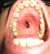 |
Fig. 1 Oroantral communication. |
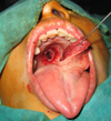 |
Fig. 2 Edge of the bank and removal of the tongue flap for a marginal pedicle. |
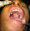 |
Fig. 3 Attachment of the flap to the palate. |
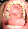 |
Fig. 4 Result after 3 months. |
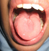 |
Fig. 5 Tongue after 3 months. |
Comments
The aim of this article was to highlight the use of a tongue flap to close an OAC. The closure of an extended OAC, especially in cases with a history of multiple procedures, is a dilemma. Moreover, palatoplasty by local flaps is unsuitable in the context of irradiated tissues.
Many authors have proposed different techniques. Locoregional flaps, such as the buccinator flap, usually with an upper pedicle, are used for closing OACs. Musculomucosal flaps receive blood supply from the facial artery, and nasal flaps received blood supply from the facial artery. The disadvantage with these flaps is the need for the extraction of a molar [3]. The temporalis muscle flap allows for the reconstruction of a wider palate [4].
The use of revascularized flaps such as prebrachial [5], an iliac ridge, and a fasciocutaneous flaps has also been reported. These flaps are indicated when the gap is greater, often resulting from tumor resections or trauma [4].
Tongue flaps were described nearly a century ago by Guerrero-Santos, and were modified by Bakamjian and Gosset [2].
Their advantages are simplicity, efficiency, and reliability, thanks to the vascularity of the tongue, which is supplied by the two lingual arteries, which are branches of the carotid arteries, on either side. In addition, there is no morbidity at the donor site. Each lingual artery has two main branches: one dorsal and the other ventral (the ranine artery). The two dorsal arteries anastomose at the proximal part of the mobile tongue , and the two ventral arteries anastomose at the distal part of the tongue (Fig. 6).
This double vascularization allows different types of tongue flaps [6]: dorsal, ventral, marginal to distal pedicle, marginal pedicle and distal bipedicle (similar to the head of hammerhead sharks).
The lingual flap may be a distal pedicle flap vascularized by the anastomosis of the ventral arteries or a proximal pedicle vascularized by the anastomosis of the dorsal arteries. The latter is not used often because of the risk of the pedicle tearing resulting from the pull from the base of the tongue.
The disadvantages of this technique are the following: two separate surgical procedures, discomfort lasting approximately 3 weeks because of LOM, reduction of lingual mobility, speech problems, and the need for nasogastric intubation in the period between the two procedures (as recommended by some practitioners).
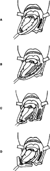 |
Fig. 6 (A) lingual vascularization according to Bracka; (B) marginolingual flap with distal pedicle; (C) marginolingual to proximal pedicle flaps; (D) distal marginolingual bipedicle flap. |
Conclusion
Tongue flap is simple and reproducible. It allows the closure of extended OACs in patients who have had multiple surgeries. It is a surgical alternative to obturator prostheses, which are often not well-tolerated by patients in the long-term. The functional and aesthetic results of tongue flaps are excellent, and the surgical procedure is simple.
Conflict of interest
The authors declare that they have no conflicts of interest in relation to this article
References
- Smith JC, Kacker A, Anand VK. Midline nasal and hard palate destruction in cocaine's abusers and cocaine’s role in rhinologic practice. Ear Nose Throat J 2002;81:172–177. [PubMed] [Google Scholar]
- Gosserez M, Stricker M, Flot F, Gola R, Malka G. Les indications du lambeau de langue dans la réparation des pertes de substance buccale. J F ORL 1973;22:921–923. [Google Scholar]
- Noboru O, Takuya O, Yoshinori I. Use of free conchal cartilage graftfor closure of a palatal fistula: an experimental study and clinicalapplication. Plast Reconstr Surg 1993;91:433–440. [CrossRef] [PubMed] [Google Scholar]
- Bettega G. La reconstruction du voile du palais. Rev Stomatol, Chir Maxillo-fac Chir Orale 2013;114:24–33. [Google Scholar]
- Macleod AM, Morrison WA, McCann JJ, Thislethwaite S, Vanderkolk CA, Ryan AD. The free radial forearm with and without bone for closure of large palatal fistulae. Br J Plast Surg 1987;40:391–395. [CrossRef] [PubMed] [Google Scholar]
- Bracka A. The blood supply of dorsal tongue flaps. Br J Plast Surg 1981;34:379–384. [CrossRef] [PubMed] [Google Scholar]
All Figures
|
Fig. 1 Oroantral communication. |
| In the text | |
|
Fig. 2 Edge of the bank and removal of the tongue flap for a marginal pedicle. |
| In the text | |
|
Fig. 3 Attachment of the flap to the palate. |
| In the text | |
|
Fig. 4 Result after 3 months. |
| In the text | |
|
Fig. 5 Tongue after 3 months. |
| In the text | |
|
Fig. 6 (A) lingual vascularization according to Bracka; (B) marginolingual flap with distal pedicle; (C) marginolingual to proximal pedicle flaps; (D) distal marginolingual bipedicle flap. |
| In the text | |
Current usage metrics show cumulative count of Article Views (full-text article views including HTML views, PDF and ePub downloads, according to the available data) and Abstracts Views on Vision4Press platform.
Data correspond to usage on the plateform after 2015. The current usage metrics is available 48-96 hours after online publication and is updated daily on week days.
Initial download of the metrics may take a while.