Fig. 2
Download original image
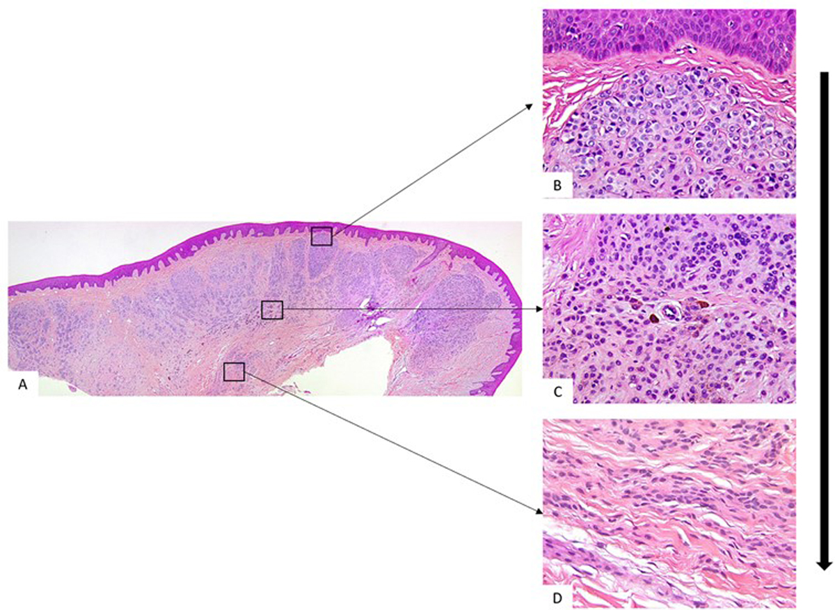
Photomicrograph of case 2 shows an intradermal nevus at low magnification (Fig. 3A, HE × 40). The architectural and cytological maturation gradient was evident at higher magnification with superficial nevus cells in theque formation without intracellular melanin (3B, HES × 100), cells in the middle zone, with only melanin in some isolated nevus cells (3C, HES × 100) and deeper nevus cells appear elongated and spindle shaped without intracellular melanin (3D, HES × 100).
Current usage metrics show cumulative count of Article Views (full-text article views including HTML views, PDF and ePub downloads, according to the available data) and Abstracts Views on Vision4Press platform.
Data correspond to usage on the plateform after 2015. The current usage metrics is available 48-96 hours after online publication and is updated daily on week days.
Initial download of the metrics may take a while.