| Issue |
J Oral Med Oral Surg
Volume 26, Number 4, 2020
|
|
|---|---|---|
| Article Number | 36 | |
| Number of page(s) | 2 | |
| Section | Cas clinique / Short case report | |
| DOI | https://doi.org/10.1051/mbcb/2020026 | |
| Published online | 26 August 2020 | |
Short Case Report
Rare case of recurrent hair in the floor of the mouth
BDF Hospital, Riffa, Kingdom of Bahrain
* Correspondence: drshilent@gmail.com
Received:
12
April
2020
Accepted:
18
June
2020
Introduction: Here we report an unusual case of recurrent hair growth in Wharton's duct in an adult male and there by exploring possible cause of it. Observation: The patient presented with recurrent hair growth in the floor of the mouth. The hair growth occurred at the right submandibular duct opening on all occasions. The patient underwent CT scan of the salivary glands which showed one stone approximately 3mm at the site which was removed under local anesthesia. Commentary: Recurrent hair growth in the floor of the mouth is rare incident. Although multiple etiologies have been described in the literature, we initially thought of the retrograde theory as patient had beard but concluded that it could be due to heterotopia as to be the possible cause of it.
Key words: dermoid cyst / oral cavity / sialolithiasis / submandibular duct / whartons duct
© The authors, 2020
 This is an Open Access article distributed under the terms of the Creative Commons Attribution License (https://creativecommons.org/licenses/by/4.0), which permits unrestricted use, distribution, and reproduction in any medium, provided the original work is properly cited.
Observation
Although hair growth in the oral cavity has been described previously here we report an unusual case of recurrent hair growth in Wharton's duct in an adult male.
A 33-year old male presented to the outpatient ENT clinic with a history of hair growth in the floor of the mouth, which he noticed five days prior to presentation. He noticed it in the mirror when he felt a foreign body sensation in the floor of the mouth. He had minimal pain at the site. On clinical examination the patient had an isolated hair fiber in the floor of the mouth, which seems to arise from the right submandibular duct opening with surrounding edema and erythema (Figs. 1 and 2). The hair fiber was removed with forceps in clinic. Swelling and pain at the site subsided within two days. Patient re visited the clinic after 10 days with recurrence of isolated hair growth at the same site.
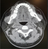
The patient underwent CT scan of the salivary glands. One stone approximately 3 mm in size was seen along the course of right submandibular duct distal to its opening in the floor of mouth. The submandibular and parotid glands appeared normal (Fig. 3).
The patient underwent removal of the hair fiber under local anesthesia in the outpatient clinic. This was done under microscopic vision using crocodile forceps. An isolated hair fiber with surrounding calcification was removed and sent for histopathology. This was followed by sialoendoscopy to further assess the submandibular duct and to remove any residual stone. Patient was kept on oral antibiotic and analgesics after procedure.
Since the removed specimen consisted of a single hair fiber with surrounding calcification the pathologist could not stain and report the findings.
On follow up the patient stated that he had a similar hair growth 20 days after the clinic procedure of removing the hair fiber from the submandibular duct. The hair growth recurred at the same site, the right submandibular duct opening and this time was removed by the patient. The patient did not have any recurrence on subsequent follow up.
 |
Fig. 1 Hair growth in the right submandibular duct near orifice. |
 |
Fig. 2 Closer view of hair growth in mouth. |
 |
Fig. 3 CT scan showing right submandibular duct stone around 3 mm near orifice. |
Commentary
Hair growth in Wharton's duct and acting as nidus for sialolith formation is a rare entity. On conducting a literature search about the possible causes of this hair growth, we found a retrograde theory in sialolithiasis formation [1]. Since our patient had facial hair growth it is possible that a facial hair fiber got trapped in the submandibular duct opening and this foreign body acted as a nidus for sialolith formation. This theory of retrograde sialolithiasis formation after hair care cannot explain the recurrence of hair growth after hair removal.
Another possibility is that the patient had a dermoid containing hair in the submandibular duct. Dermoid in the floor of mouth is a well known entity [2], but in our case there was no cystic lesion surrounding the hair fiber in the floor of the mouth. And recurrent growth of hair cannot be explained by this theory.
Isolated hair growth in the oral cavity mucosa which can be explained by another entity heterotopia reported in the literature [3,4] but this aberrance of hair growth has never been reported in the floor of the mouth to the best of the author's knowledge.
In this case the repeated occurrence can be due to insufficient removal of hair bulb but second time we tried our best to completely remove the hair bulb still there was recurrence after 20 days.
Sialometaplasia of floor of oral cavity as described in literature [5] could be the etiology but patient had little or no pain at the site, presence of hair and absence of necrotizing ulcer does not much support this being the cause.
Although multiple etiologies have been described in the literature, we would agree on the cause could be due to heterotopia which led to aberrant growth of hair in the floor of mouth.
Conflicts of interests
The authors declare that they have no conflicts of interest in relation to the publication of this article.
References
- Marchal F, Kurt AM, Dulguerov P, Lehmann W. Retrograde theory in sialolithiasis formation. Arch Otolaryngol Head Neck Surg 2001;127:66–68. [CrossRef] [PubMed] [Google Scholar]
- Jadwani S, Misra B, Kallianpur S, Bansod S. Dermoid cyst of the floor of the mouth with abundant hair: a case report. J Maxillofac Oral Surg 2009;8:388–389. [PubMed] [Google Scholar]
- Agha-Hosseini F, Etesam F, Rohani B. A boy with oral hair: case report. Med Oral Patol Oral Cir Bucal 2007;12:357–359. [Google Scholar]
- Rochefort J, Guedj A, Lescaille G, Hervé G, Agbo-Godeau S. A hair on the tongue. Rev Stomatol Chir Maxillofac Chir Orale 2016;117:357–358. [PubMed] [Google Scholar]
- Alande C, Fenelon M, Catros S, Fricain JC. Persistent ulceration of the oral floor: a case of necrotizing sialometaplasia of the sublingual gland? Med Bucc Chir Bucc 2017;23:188–189. [CrossRef] [Google Scholar]
All Figures
 |
Fig. 1 Hair growth in the right submandibular duct near orifice. |
| In the text | |
 |
Fig. 2 Closer view of hair growth in mouth. |
| In the text | |
 |
Fig. 3 CT scan showing right submandibular duct stone around 3 mm near orifice. |
| In the text | |
Current usage metrics show cumulative count of Article Views (full-text article views including HTML views, PDF and ePub downloads, according to the available data) and Abstracts Views on Vision4Press platform.
Data correspond to usage on the plateform after 2015. The current usage metrics is available 48-96 hours after online publication and is updated daily on week days.
Initial download of the metrics may take a while.


