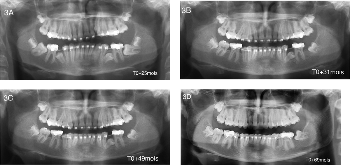
Fig. 3
Download original image
Patient B; 4 years after radiological follow-up. (A) First panoramic X-ray (T0 + 25 months). The start of orthodontic treatment with delayed eruption of teeth 35, 45, and 37 tilted on the mesial side. Four wisdom teeth were unerupted. (B) Panoramic X-ray after 6 months of follow-up (T0 + 31 months). Tooth 37 was extracted. (C) Panoramic X-ray after 2 years of follow-up (T0 + 49 months). Tooth 45 was in a normal position, and tooth 35 was still unerupted. (D) Panoramic X-ray after approximately 3.5 years of follow-up (T0 + 69 months). Tooth 35 was in a normal position, and tooth 38 was surgically exposed for orthodontic procedures. Teeth 45 and 35 show a radicular hook apical region.
Current usage metrics show cumulative count of Article Views (full-text article views including HTML views, PDF and ePub downloads, according to the available data) and Abstracts Views on Vision4Press platform.
Data correspond to usage on the plateform after 2015. The current usage metrics is available 48-96 hours after online publication and is updated daily on week days.
Initial download of the metrics may take a while.