Fig. 2
Download original image
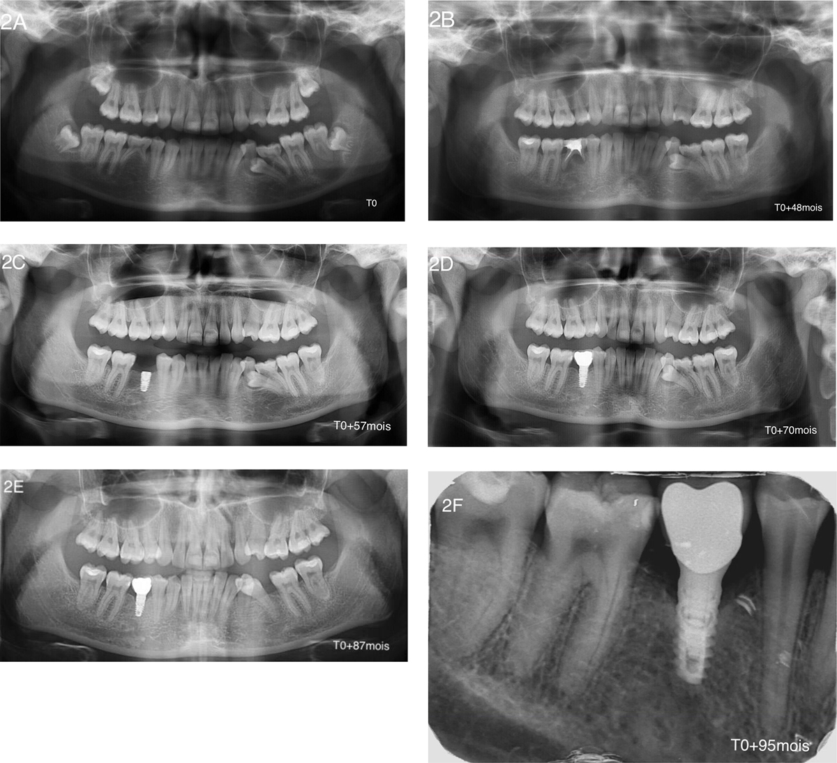
Patient A; 8 years after radiological follow-up. (A) Initial panoramic X-ray. Agenesis of the upper first premolars and second lower right premolar, delayed eruption of tooth 35, remaining teeth 75 and 85, unerupted 4 wisdom teeth, and follicular cyst involving tooth 38. (B) Panoramic X-ray after 48 months of follow-up. Tooth 85 presented a periapical lesion. This chronic infection required tooth extraction. (C) Panoramic X-ray after 57 months of follow-up. Two months post-implantation (XiveS*, 3,8*8 mm, Dentsply-Sirona, Manheim, Germany) for replacement of tooth 45. (D) Panoramic X-ray after 70 months of follow-up (1.5 years after implantation). Peri-implant bone level stability and successful osteointegration. (E) Panoramic X-ray after 87 months of follow-up (29 months after implantation). Peri-implant bone level stability and successful osteointegration. Tooth 35 was erupting in good position. (F) Retroalveolar X-ray after 95 months of follow-up for implant 45 (3 years post-implantation). Peri-implant bone level stability under the level of the first thread.
Current usage metrics show cumulative count of Article Views (full-text article views including HTML views, PDF and ePub downloads, according to the available data) and Abstracts Views on Vision4Press platform.
Data correspond to usage on the plateform after 2015. The current usage metrics is available 48-96 hours after online publication and is updated daily on week days.
Initial download of the metrics may take a while.