| Issue |
J Oral Med Oral Surg
Volume 27, Number 4, 2021
|
|
|---|---|---|
| Article Number | 56 | |
| Number of page(s) | 6 | |
| DOI | https://doi.org/10.1051/mbcb/2021023 | |
| Published online | 25 November 2021 | |
Case Report
Interest and outcomes of alveolar distraction osteogenesis for dental implant rehabilitation following mandibular reconstruction
1
Université de Reims Champagne Ardenne, Faculté de Médecine, Reims 51100, France
2
Service de chirurgie maxillo-faciale et stomatologie, Hôpital de la Pitié Salpêtrière, 75013 Paris, France
3
Université de Paris-Sorbonne, Faculté de Médecine, 75006 Paris, France
* Correspondence: This email address is being protected from spambots. You need JavaScript enabled to view it.
Received:
6
November
2020
Accepted:
15
April
2021
Abstract
Introduction: Alveolar distraction osteogenesis (ADO) is one of vertical bone augmentation technique used to restore the vertical bone discrepancy between the transplanted graft and the residual alveolar bone after mandibular reconstruction. The aim of this article is to present the clinical outcomes of ADO applied to reconstructed mandible for three patients. Observation: Three patients underwent alveolar distraction osteogenesis procedure on mandibula reconstructed by fibula free flap (n = 2) or iliac crest free flap (n = 1). The mean bone height increase was 11 mm. 16 implants have been placed in all patients, with a success rate of 100% and a mean follow-up of 25 months. Two complications occurred without compromising the final outcome of the procedure: one fracture of the basal bone and one bony interference, both during activation phase. Commentaries: ADO can be performed on reconstructed mandible with free flap to restore alveolar height. An acceptable implant-to-crown ratio allows an optimal supported implant prosthesis. The particular antecedent of our patients can lead to uncommon complications such as basal bone fracture, but doesn't seem to compromise implant placement with good success rate. Conclusions: ADO performed on reconstructed mandible allows appropriate dental implant rehabilitation, achieving good esthetics and occlusal outcomes.
Key words: Alveolar distraction / dental implant / mandibular reconstruction / free flap
© The authors, 2021
 This is an Open Access article distributed under the terms of the Creative Commons Attribution License (https://creativecommons.org/licenses/by/4.0), which permits unrestricted use, distribution, and reproduction in any medium, provided the original work is properly cited.
Introduction
Vascularized free flap has become an indispensable modality for aesthetical and functional reconstruction for mandibular defects [1]. However, the insufficient height of transplanted free flap leads to an increased interarch distance with unfavorable intermaxillary relationship, thus, unsatisfying prosthetic results. An unfavorable implant-to-crown ratio can generate an overload on dental implants [2], implant fracture, prosthetic components damages, aesthetic problems and difficulties in maintaining oral hygiene [3].
An alveolar vertical augmentation could prevent these outcomes, and several methods exist such as Guided Bone Regeneration (GBR), autologous bone blocks, osteotomy and alveolar distraction osteogenesis (ADO) [4]. Using autologous bone graft technique for severe vertical bone atrophy leads to higher risk of wound dehiscence with bone graft loss due to soft tissue defect. ADO, defined by newly bone formation between two divided bone segments gradually distracted with a mechanical device [5], presents the advantage to expand soft tissue at the same time [5], and to reduce postoperative morbidity (no need of bone harvesting) [6].
The concept of ADO was introduced by Codivilla in 1905 [7]. Its first application was for orthopedic deformities of long bone [8] by Ilizarov in 1954. Finally, Mac Carthy et al. [9], in 1992, used it in maxillo-facial surgery.
This cases series presents the outcomes of ADO in reconstructed mandibles for dental implant rehabilitation.
Observation (Tab. I)
From October 2016 to May 2019, three ADO was performed in 3 male patients, 23–28 years old (mean 25 years old). They were previously treated for ameloblastoma (n = 1) and osteosarcoma (n = 2) using interruptive mandibulectomy reconstructed with fibular free flap (n = 2) or iliac free flap (n = 1). Patients' sex and age at the time of the distractor implantation are presented in Table I, just like the intervals between transplantation and start of ADO.
Oral evaluation objectives an important vertical ridge discrepancy (ranging from 15 mm to 20mm) with insufficient amount of keratinized gingiva of the reconstructed site (Fig. 1).
Residual bone height estimated by Cone Beam CT was satisfying (mean of 12 mm) (Fig. 2), but the implant-to-crown ratio was heightened (>1:3) (Fig. 3).
To reduce the prosthetic space for appropriate implant rehabilitation, the same ADO protocol was applied to all three. The procedure differed only in the locations of the affected mandibular segments. These locations are listed in Table II.
The procedure (Fig. 4) was carried out under general anesthesia with nasotracheal intubation. The patients were placed in the supine position, cutaneous and intra-oral disinfection with povidone-iodine and surgical draping were performed.
A buccal incision was made and full thickness flap was elevated to visualize the distraction site and anatomical obstacles. The lingual mucoperiosteal attachment is preserved to guarantee the vascularization of the future transport fragment (Fig. 4a).
A mandibular distractor (KLS Martin), shaped to the planed site using 3D printed mandible before surgery, is fixed with screws (∅1.5 mm × 5 mm screw for the transport fragment and ∅2mm bi cortical screw for basal bone).
After pre drilling stage, two divergent verticals and one horizontal osteotomy are performed using PiezoSurgery (Fig. 4b), and the distractor is removed to finalize the osteotomy (Fig. 4c).
After mobilizing the transport bone fragment (Fig. 4d), the distractor was fixed into its initial position (Fig. 4e).
The device was activated to control the absence of bony interferences, and evaluate the distraction vector.
The flap was closed in one layer with Vicryl 3.0.
All patients received oral antibiotics for 7 days, non-steroidal analgesics post-operatively and had appropriate oral hygiene with 0.2% chlorhexidine mouth rinse. Postoperative instructions included a soft diet for 2 week, and removable prostheses were not allowed.
Description of the population.
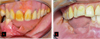 |
Fig. 1 Important vertical ridge discrepancy (ranging to 15 mm to 20mm) with insufficient amount of keratinized mucosa after a segmental mandibulectomy reconstructed with fibular free flap (A: Patient n°1) (B: Patient n°2). |
 |
Fig. 2 Residual bone height mean of 12mm (A: patient n°1), 15mm (B: patient n°2), and 12mm (C: patient n°3). |
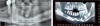 |
Fig. 3 Implant −to-crown ratio superior to 1 for 3(A: patient n°1, B: patient n°2). A minimum bone gain of 10mm would optimize the supported implant prosthesis by reducing this ratio. |
Data on resection, distraction, implants and prosthetic solution in three patients of the study.
 |
Fig. 4 (a) Buccal incision − elevation of a full thickness vestibular flap with preservation of lingual mucoperiosteal attachment. (b) Two divergent vertical and one horizontal osteotomy are performed using Piezosurgery. (c) Distractor is removed to finalize the osteotomies. (d) Transport bone fragment is mobilized. (e) Distractor fixed into his initial position. |
Latency phase
A five days period is required to obtain primary wound closure and to transform the blood clot trapped in the gap into rich collagen tissue [10].
Activation phase
Depending on the desired bone height, distraction at 1 mm per day (0.5 mm × 2) [11] was applied for periods varying between 12 and 15 days. The mechanical traction applied on the callus stimulates its transformation into newly bone tissue [10].
Consolidation phase
The fragment is stabilized into its final position by the rigid device for 12 weeks, leading to the mineralization of the callus present in the distraction gap [10].
Implant placement
At the end of the consolidation period, all patients received endosseous implants using surgical guide under local anesthesia (Fig. 5). The distraction device was removed at the same time. Correction of the vestibular soft tissue could also be performed at this stage if necessary. An orthopantomogram was realized after surgery to control the implant placement (Fig. 5).
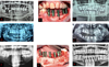 |
Fig. 5 Significant vertical ridge augmentation allowing implant placement after distractor removal for patient n°1 (Figs 5A, 5B, 5C), for patient n°2 (Figs 5D, 5E) and for patient n°3 (Figs 5F, 5G, 5H). |
Results
Bone volume estimated by cone beam CT (CBCT) at the end of consolidation phase quantified a mean vertical ridge augmentation of 13.6 mm (Tab. I), allowing appropriate implant placement for all patients.
One fracture of the basal bone (Fig. 6a) on either side of the distractor device was reported for one patient few days after activation. A new distraction device was placed with longer basilar plate to stabilize the fracture site at the same time (Fig. 6b).
A premature stop of distracted fragment due to bony interference occurred five days before the end of activation phase for one patient, ending the ADO protocol with a 15 mm bone gain.
Despite these complications, the vertical correction of the defect was satisfying.
16 implants have been placed in the distraction area. At one-year follow-up for one patient and two-years for the two others, an implant success rate of 100% according to Albrektsson's criteria [12] was recorded.
An acceptable crown/implant index was restored. As a result, a convenient implant supported prothesis was achieved, improving orofacial function (mastication, phonation, salivatory capacity) and oral hygiene maintenance.
 |
Fig. 6 Fracture of the basal bone on either side of the distraction site (6a), fixed with a longer basal plate of a new distractor device (6b). |
Discussion
Implant supported prosthetic rehabilitation of patients with severe bone atrophy after tumor resection remains a challenge, mostly because of insufficient height of the free flap used for mandible reconstruction.
A stable and significant increase of vertical bone height using ADO on free flap permitted an adapted implant rehabilitation [13]. The distraction procedure has also significantly improved the alveolar ridge anatomy and the formation of soft tissue in the vestibulum of the affected region. A vestibuloplasty could have been performed in the same time as implant placement but wasn't necesseray in those cases.
In the present study, the follow-up period immediately after implant insertion ranged from 12 to 25 months (average 20 months). The overall implant success rate in our study was 100% (16 of 16), which is close to the results found in the literature (100% of survival rate and 84% of success rate according to Wang et al. [3]).
The mean bone loss around the implant, measured on the orthopantomograms, at one year follow up was low (<1 mm). However, the use of more precise technique such as long-cone technique and intra-oral films should be preferable then panoramic radiograph for implant follow-up. A clinical study of 5 cases of ADO on reconstructed mandible conducted by Cheung et al. [14] in 2013 reported similar results, with a minimal resorption rate ranging to 0,5 mm to 1,0 mm at 1 one year after implantation with an implant success rate of 100%.
Lizio et al. [2] reported an implant success rate of 89% and a mean bone resorption of 2,5 mm with a mean follow up of 38 months. Kunkel et al. [15] reported similar results with an implant survival rate of 90% with a mean follow up of 39 months.
One fracture of the basal fibular cortex occurred but this complication seems to remain relatively rare for non-reconstructed mandible as reported by Zhao and Al [8,16]) (4 basal bone fracture on 353 ADO). It is commonly related to the mechanical weakness of the residual basal bone [17].
A premature stop of the distraction device for one patient leaded to a 15 mm bone height instead of the 20 mm expected. Knowing that iliac crest free flap owns a higher residual bone than fibular free flap [17], the resulting implant-to-crown ratio was still satisfying allowing correct implant placement. Slightly divergent vertical osteotomy [15], done in these cases, is supposed to minimize the occurrence of bony inhdrance.
Conclusion
Despite the occurrence of complications, the satisfying results of our study support the use of ADO for the rehabilitation of mandibular defects following tumor, but a larger number of patients and a longer follow up period should be considered to improve the power of our study.
Conflicts of interests
The authors declare that they have no conflict of interest in this study.
Informed consent
The author declare that informed consent not required.
Ethical committee approval
The author declare that Ethical approval not required.
Source of funding
This research did not receive any specific funding.
References
- A Comparison of Bone Resorption Over Time: An Analysis of the Free Scapular, Iliac Crest, and Fibular Microvascular Flaps in Mandibular Reconstruction − PubMed [Internet]. [cité 23 sept 2020]. Disponible sur: https://pubmed.ncbi.nlm.nih.gov/27725102/ [Google Scholar]
- Lizio G, Corinaldesi G, Pieri F, Marchetti C. Problems with dental implants that were placed on vertically distracted fibular free flaps after resection: a report of six cases. Br J Oral Maxillofac Surg 2009;47:455–460. [CrossRef] [PubMed] [Google Scholar]
- Wang F, Wu Y, Zhang C, Zhang Z. Dental implant performance in vertically distracted fibular grafts after mandibular reconstruction: a pilot series of 12 patients. Int J Oral Maxillofac Implants 2013;28:1311–1321. [CrossRef] [PubMed] [Google Scholar]
- Hameed MH, Gul M, Ghafoor R, Khan FR. Vertical ridge gain with various bone augmentation techniques: a systematic review and meta-analysis. J Prosthodont Off J Am Coll Prosthodont 2019;28:421–427. [CrossRef] [PubMed] [Google Scholar]
- Cheung LK, Hariri F, Chua HDP. Alveolar distraction osteogenesis for oral rehabilitation in reconstructed jaws. Curr Opin Otolaryngol Head Neck Surg 2011;19:312–316. [CrossRef] [PubMed] [Google Scholar]
- Chiapasco M, Zaniboni M, Rimondini L. Autogenous onlay bone grafts vs. alveolar distraction osteogenesis for the correction of vertically deficient edentulous ridges: a 2-4-year prospective study on humans. Clin Oral Implants Res 2007;18:432–440. [CrossRef] [PubMed] [Google Scholar]
- Codivilla A. The classic: on the means of lengthening, in the lower limbs, the muscles and tissues which are shortened through deformity. Clin Orthop 2008;466:2903–2909. [CrossRef] [PubMed] [Google Scholar]
- Ilizarov GA. The principles of the Ilizarov method. Bull Hosp Jt Dis Orthop Inst 1988;48:1–11. [PubMed] [Google Scholar]
- McCarthy JG, Schreiber J, Karp N, Thorne CH, Grayson BH. Lengthening the human mandible by gradual distraction. Plast Reconstr Surg 1992;89:1–8; discussion 9–10. [CrossRef] [PubMed] [Google Scholar]
- Natu SS, Ali I, Alam S, Giri KY, Agarwal A, Kulkarni VA. The biology of distraction osteogenesis for correction of mandibular and craniomaxillofacial defects: a review. Dent Res J 2014;11:16–26. [PubMed] [Google Scholar]
- Ilizarov GA. The tension-stress effect on the genesis and growth of tissues. Part I. The influence of stability of fixation and soft-tissue preservation. Clin Orthop 1989;238:249–281. [CrossRef] [Google Scholar]
- Albrektsson T, Zarb G, Worthington P, Eriksson AR. The long-term efficacy of currently used dental implants: a review and proposed criteria of success. Int J Oral Maxillofac Implants 1986;1:11–25. [PubMed] [Google Scholar]
- Dholam KP, Bachher GK, Yadav PS, Quazi GA, Pusalkar HA. Assessment of quality of life after implant-retained prosthetically reconstructed maxillae and mandibles postcancer treatments. Implant Dent 2011;20:85–94. [CrossRef] [PubMed] [Google Scholar]
- Cheung LK, Chua HDP, Hariri F, Pow EHN, Zheng L. Alveolar distraction osteogenesis for dental implant rehabilitation following fibular reconstruction: a case series. J Oral Maxillofac Surg Off J Am Assoc Oral Maxillofac Surg 2013;71:255–271. [CrossRef] [Google Scholar]
- Kunkel M, Wahlmann U, Reichert TE, Wegener J, Wagner W. Reconstruction of mandibular defects following tumor ablation by vertical distraction osteogenesis using intraosseous distraction devices. Clin Oral Implants Res 2005;16:89–97. [Google Scholar]
- Zhao K, Wang F, Huang W, Wu Y. Clinical outcomes of vertical distraction osteogenesis for dental implantation: a systematic review and meta-analysis. Int J Oral Maxillofac Implants 2018;33:549–564. [CrossRef] [PubMed] [Google Scholar]
- Makiguchi T, Yokoo S, Hashikawa K, Miyazaki H, Terashi H. Evaluation of bone height of the free fibula flap in mandible reconstruction. J Craniofac Surg 2015;26:673–676. [CrossRef] [PubMed] [Google Scholar]
All Tables
Data on resection, distraction, implants and prosthetic solution in three patients of the study.
All Figures
|
Fig. 1 Important vertical ridge discrepancy (ranging to 15 mm to 20mm) with insufficient amount of keratinized mucosa after a segmental mandibulectomy reconstructed with fibular free flap (A: Patient n°1) (B: Patient n°2). |
| In the text | |
|
Fig. 2 Residual bone height mean of 12mm (A: patient n°1), 15mm (B: patient n°2), and 12mm (C: patient n°3). |
| In the text | |
|
Fig. 3 Implant −to-crown ratio superior to 1 for 3(A: patient n°1, B: patient n°2). A minimum bone gain of 10mm would optimize the supported implant prosthesis by reducing this ratio. |
| In the text | |
|
Fig. 4 (a) Buccal incision − elevation of a full thickness vestibular flap with preservation of lingual mucoperiosteal attachment. (b) Two divergent vertical and one horizontal osteotomy are performed using Piezosurgery. (c) Distractor is removed to finalize the osteotomies. (d) Transport bone fragment is mobilized. (e) Distractor fixed into his initial position. |
| In the text | |
|
Fig. 5 Significant vertical ridge augmentation allowing implant placement after distractor removal for patient n°1 (Figs 5A, 5B, 5C), for patient n°2 (Figs 5D, 5E) and for patient n°3 (Figs 5F, 5G, 5H). |
| In the text | |
|
Fig. 6 Fracture of the basal bone on either side of the distraction site (6a), fixed with a longer basal plate of a new distractor device (6b). |
| In the text | |
Current usage metrics show cumulative count of Article Views (full-text article views including HTML views, PDF and ePub downloads, according to the available data) and Abstracts Views on Vision4Press platform.
Data correspond to usage on the plateform after 2015. The current usage metrics is available 48-96 hours after online publication and is updated daily on week days.
Initial download of the metrics may take a while.