Fig. 2
Download original image
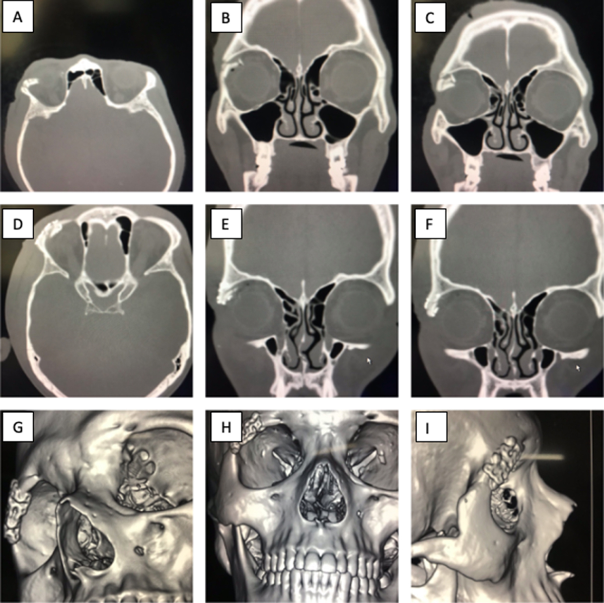
Preoperative axial (A) and coronal (B and C) sections showing comminuted fracture of the right lateral orbital rim. Postoperative axial (D) and coronal (E and F) sections showing accurate reduction of right lateral rim fragments. Postoperative 3D reformatted images (G, H and I) showing the titanium mesh shaped and adapted to the right lateral orbital rim and cupping the minute bone fragments in shape.
Current usage metrics show cumulative count of Article Views (full-text article views including HTML views, PDF and ePub downloads, according to the available data) and Abstracts Views on Vision4Press platform.
Data correspond to usage on the plateform after 2015. The current usage metrics is available 48-96 hours after online publication and is updated daily on week days.
Initial download of the metrics may take a while.