| Issue |
J Oral Med Oral Surg
Volume 27, Number 3, 2021
|
|
|---|---|---|
| Article Number | 43 | |
| Number of page(s) | 6 | |
| DOI | https://doi.org/10.1051/mbcb/2021021 | |
| Published online | 03 September 2021 | |
Original Research Article
Significance of cervical node necrosis in preoperative MRI as a prognostic indicator: retrospective study of patients with SCC of tongue
Kidwai Memorial Institute of Oncology, Bangalore, India
* Correspondence: This email address is being protected from spambots. You need JavaScript enabled to view it.
Received:
24
November
2020
Accepted:
16
May
2021
Abstract
Aim: To ascertain the prognostic value of cervical nodal necrosis (CNN) observed in patients of tongue squamous cell carcinoma with magnetic resonance imaging. Materials and methods: In this retrospective observational study, records of 144 patients diagnosed with newly diagnosed SCC of tongue were considered. Preoperative MRI study, demographic and clinical data were reviewed. Based on MRI reports, patients were categorised into: with or without the presence of cervical node necrosis (CNN or non CNN). Subsequent treatments, histopathological reports and follow up data were studied to determine key prognostic elements, overall survival and disease free survival by statistical analysis. Results: The incidence of CNN was 55.55% in the study sample. CNN category, depth of invasion, N stage and extra nodal extension were significant negative prognostic factors for overall and disease free survival. Conclusion: Based on our results, pre operative MRI based presence of cervical node necrosis in tongue squamous cell carcinoma is an independent prognostic indicator for poor overall and disease free survival. Long term prospective studies with larger cohorts could be undertaken to establish its role as an important biomarker for precision treatments.
Key words: Cervical node necrosis / MRI / squamous cell carcinoma / tongue / prognostic factor
© The authors, 2021
 This is an Open Access article distributed under the terms of the Creative Commons Attribution License (https://creativecommons.org/licenses/by/4.0), which permits unrestricted use, distribution, and reproduction in any medium, provided the original work is properly cited.
Introduction
The Indian subcontinent has one of the highest burdens of oral malignancy in the world [1]. Even though the gingivobuccal cancer is termed as the Indian cancer, tongue cancer also presents itself frequently [2]. The tongue, because of its anatomical characteristics of muscle bundles and rich lymphovascular network, has a higher risk of cervical metastasis [3]. It is well known that the presence of lymph node metastasis is the most vital prognostic indicator, affecting overall and locoregional rates and thus its Accurate assessment is critical for evaluating treatment options and predicting prognosis in oral cancer patients [4]. Consequently, even in tongue cancer patients exhibiting clinically negative nodal disease, more than 30% of them will have cervical metastasis [5]. Thus the AJCC staging in its 8th edition has highlighted a nodal characteristic (ENE-Extracapsular spread) and a tumour characteristic (DOI − depth of invasion) as additional staging parameters affecting patient outcomes [6]. Since then there has been robust research in the field of oncology to identify patient characteristics, imaging features, blood parameters, histopathological criteria, molecular biomarkers in order to predict the prognosis of the disease.
Most patients undergo clinical, imaging and histopathological evaluation for disease diagnosis and staging. Computed tomography, Magnetic resonance imaging, Ultrasonography, Positron emission tomography are utilized as per the requirement with their corresponding sensitivity and specificity rates.
SCC of the tongue often requires MRI (with or without CT) to study tumour dimensions, its anatomical relations and cervical nodal involvement. Nodal necrosis is an important imaging feature used to distinguish between benign and malignant lymph nodes [7,8]. At magnetic resonance (MR) imaging, CNN is seen as a focal area of high signal intensity on T2-weighted images and as an area of low signal intensity on contrast material–enhanced T1-weighted images, with or without a surrounding rim of enhancement. The aim of this retrospective study was to ascertain the prognostic value of cervical node necrosis in patients with tongue SCC based on their preoperative MR findings.
Materials and methods
In this retrospective observational study at our institute (Regional cancer centre), records from January 2013 to 2015 were included. The following Inclusion criteria were considered: records of patients with clinically node positive, histopathologically proven squamous cell carcinoma of tongue who underwent primary surgery followed by adjuvant therapy when indicated. The exclusion criteria was: records of patients with N0 nodal disease, presence of distant metastasis, synchronous malignancy, recurrent tongue SCC cases. Patients underwent MRI for the evaluation of primary tumour, cervical node status and CT scan of chest to detect any pulmonary masses (distant metastases). Patient information was anonymized before analysis. Subsequently, 144 patient records (87 men and 57 women; mean age, 53.5 years; range, 31–82 years) were included in our study. Preoperative MRI images of the neck were available for all patients and CNN/ non CNN categorization was done. Data pertaining to patient demographics, the TNM classification (8th edition of the AJCC), tobacco use, and the status at the last follow-up visit (alive or dead; disease-free survival [DFS] or treatment failure [TF]) were collected from the medical records department. For this study, TF was defined as either residual disease at the end of definitive therapy, locoregional recurrence after a disease-free period, presence of distant metastases with or without recurrence or disease-related mortality. Any TF was noted by histological confirmation and imaging evidence in the follow up period. For cases without TF, the follow-up period was measured in months from the time of diagnosis. Data were censored to the last follow-up date in the medical records till December 31, 2019.
Imaging and collection
A 1.5 T magnet MRI (Ingenia, Philips) with a head and neck coil was used to perform MRI PNS and neck. As per institution protocol, Axial spin-echo (SE) T1-weighted and fat-saturated fast SE T2-weighted images were acquired. Axial, sagittal T1-weighted spin-echo and coronal T1-weighted fat-suppressed spin-echo images were sequentially obtained before and after intravenous injection of 0.1 mmol/kg body wt of gadolinium based contrast material.
A blinded consensus review of the MRI images was done by an experienced radiologist. Patients with lymph nodes showing varied morphology were grouped together and evaluated for the presence (CNN group; Fig. 1) or absence of nodal necrosis (non-CNN group). The criteria for diagnosing a cervical node necrosis in an MR image was a focal area of high signal intensity on T2-weighted images or a focal area of low signal intensity on contrast-enhanced T1-weighted images, with or without a surrounding rim of enhancement (Fig. 1).
 |
Fig. 1 Cervical nodal necrosis observed in MRI studies of a patient diagnosed with squamous cell carcinoma of tongue. |
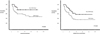 |
Fig. 2 A,B —Kaplan-Meier graph showing overall survival and disease-free survival rates of patients with tongue squamous cell carcinoma with cervical nodal necrosis (CNN group) or without CNN (non-CNN group) on preoperative MRI studies. |
Treatment and clinical follow-up
All patients underwent surgical resection with neck dissection as the primary treatment. Based on tumour location and preoperative imaging findings, modified radical or radical neck dissection on the ipsilateral side was performed for 95 patients (65.97%), and bilateral neck dissection was performed for 49 patients (34.0%). Subsequently, 120 (83.3%) of these patients had undergone adjuvant therapy i.e. radiation therapy with or without chemotherapy based on the histopathological features (RT indicated for T3 or T4 tumor; compromised surgical resection margins (<5 mm from the inked surface of the specimen); presence of lympho-vascular invasion (LVI) and/or perineural invasion (PNI); and positive lymph nodes with or without extracapsular invasion (ECE); CT indicated for microscopic positive margins and/or ECE). Standard of care RT via EBRT/ IMRT was administered. All patients were followed up for the first 1 month after established treatment culminated in the first year and every 3 months for the next 2 years till they were disease-free. Subsequent follow-ups were scheduled every 6 months for the next 2 years and annually thereafter.
Statistical analysis
The Statistical Package for the Social Sciences (SPSS, IBM) version 23.0, was used for statistical analysis. Overall survival (OS), disease-free survival (DFS) were estimated with the Kaplan-Meier method for visual comparison of OS and DFS between the CNN and non-CNN groups and were compared using a log-rank test. Overall survival (OS) was ciphered from the date of disease diagnosis to the date of the last follow-up visit or death due to disease associated reasons. Disease-free survival (DFS) was calculated from the date of treatment conclusion to the date of TF recording. Subgroup analysis via Fisher exact test and independent-samples t tests were used to compare clinical and pathologic characteristics between the CNN and non CNN groups (Tab. I). Univariate analysis to determine predictors of OS and DFS among the following variables: presence or absence of CNN on preoperative imaging, presence or absence ENE, depth of invasion, T classification and N classification was done (Tab. II). A p value of <0.05 was considered statistically significant. Kaplan–Meier graphs were plotted (Fig. 2).
Clinical and Pathologic Characteristics of Patients with SCC of Tongue With Cervical Nodal Necrosis (CNN) (CNN Group) or Without CNN (Non-CNN Group) on Preoperative MR Imaging.
Univariate Analysis of Variables Associated With Survival Outcomes of Patients With Tongue Squamous Cell Carcinoma With Cervical Nodal Necrosis (CNN) (CNN Group) or Without CNN (Non-CNN Group) on Preoperative MRI.
Results
The incidence of CNN in patients with cervical node metastases was 55.55% (80/144). The mean maximal axial diameter of lymph node metastases was 18.2 ± 1.1 (SD) mm for all patients, 27.9 ± 8.4 mm for patients with CNN, and 12.5 ± 7.5 mm (range, 3.8– 11.9 mm) for patients without CNN. 35 patients showed level I (Ia or Ib) nodal necrosis, 59 showed level II (IIa or IIb) nodal necrosis, 19 showed level III nodal necrosis, 9 showed level IV nodal necrosis, and 4 showed level V nodal necrosis. 32 patients were diagnosed with grade 1 SCC, 63 with grade 2 SCC and 49 patients with grade 3 SCC. Table I presents the patient demographic characteristics and pathologic characteristics of the tumors according to the presence of CNN. There were no significant differences in most clinical and pathologic variables between the CNN and non-CNN groups; however, the numbers of patients with advanced nodal disease, higher histopathological differentiation, and pathologically proven ENE were higher in the CNN group than in the non-CNN group. The mean follow-up duration for the total case population was 32.6 months. TF occurred in 39 patients at a mean of 12.5 months after treatment completion. Univariate analysis using a Cox proportional hazard model revealed that presence of CNN on preoperative imaging, presence of ENE, depth of invasion, T and N classification were significantly associated with OS and DFS (Tab. II). Kaplan-Meier survival curves showed that the OS and DFS rates were significantly lower in patients with CNN (p = 0.008) than in those without CNN (p = 0.013).
Discussion
Patients with head and neck cancers require precise assessment to deduce the prognosis associated and the therapeutic option to be given. Apart from the primary tumour characteristics, cervical nodes have a more significant role affecting the survival of patients. Patient morbidity and mortality in oral squamous cell carcinoma are consequential to regional and distant metastasis. The criteria suggested by Brekel et al., have been accepted as the most reliable criteria for evaluating metastases in cervical lymph nodes [9]. Various nodal parameters have been substantiated as important prognostic factors, including size, central necrosis and extracapsular extension [10]. Ding et al., demonstrated MRI diagnostic criteria of cervical lymph node metastasis include nodal size, central nodal necrosis, and irregular contour of lymph nodes [11]. High treatment failure rates in tongue scc patients despite aggressive surgical and timely adjuvant therapy have led to focussed research on understanding tumour biology, identifying patient specific prognostic markers based on hematological, radiological and histological parameters for disease stratification and delivering precision therapy. Radiological parameters could be easy and feasible options for the same [12]. Our study established the presence of CNN on preoperative imaging being significantly related to advanced disease with survival rates of patients with CNN lower than that of patients without CNN. In our study, there was a significant correlation between presence of CNN and incidence of ENE (P = 0.003), which was associated with poor overall survival (P = 0.012). Presence of central nodal necrosis (CNN) has been correlated with aggressive malignancy [13].
The histological grade was also seen to be associated with the incidence of CNN in our study (P = 0.032). This is supportive of the findings by Morimoto et al., who suggested that CNN findings may depend on the tumour differentiation [14]. The central area of low attenuation in CNN might reflect tumour infiltration, including a tumour necrosis nest and keratinization of the tumour cells. Tumour aggression characteristics such as higher grade and greater depth of invasion have been positively correlated with nodal necrosis in our study. Metastasis central necrosis is considered a biologically late event in the evolution of tumour within a lymph node. Central node necrosis may be a marker for more intrinsically aggressive disease, irrespective of whether microscopic or macroscopic ECS is present [12].
The Kaplan Meier graph clearly indicated the significant poorer outcomes ( both overall and disease free survival) in patients with CNN when compared to those without nodal necrosis. The association between CNN and post operative ENE was significant in our study. This was consistent with other studies by Zoumalan et al. Randall et al. and Baik et al. who established that CNN visible on preoperative imaging was a useful indicator of extracapsular nodal spread and hence impaired overall survival [12,15–17]. Myer et al. in their study associated poor outcomes and poorer prognosis in patients with presence of extra capsular spread [18].
It has been noted that extracapsular spread can occur even at the early stages of tumour growth and nodal involvement [19]. This is attributed to micrometastasis which are not detected in imaging studies. ENE can occur irrespective of tumour or nodal size which has been similarly noted with nodal necrosis in our study. In their paper on establishing a nodal score for patients with oral SCC, Alkulaibi et al. proposed a higher score for nodes with central necrosis as an indication of aggressive disease [20]. Necrosis and tumour hypoxia are coexistent in tumour biology and this has been singled out as a major cause of radiation resistance.
Baik et al. and Lan et al. had highlighted the prognostic value of cervical node necrosis in CT and MRI studies [16,21]. In our analysis, the survival outcomes in the CNN group were critically poorer than those in the non-CNN group and similar to those of patients with higher N stage disease without CNN.
The retrospective nature of the study poses a limitation as large prospective cohort studies could be taken up to assess the severity of nodal necrosis and highlight its value as a reliable prognostic marker. The difference in the dosage of radiation given to both the CNN and non CNN groups could also be correlated. Other hematological parameters such as lymphocyte neutrophil ratio, hypoxia dependent factor, VEGF etc. could also be studied in correlation to cervical node necrosis.
Conclusion
In conclusion, CNN is seen to be a promising independent negative prognostic factor in tongue squamous cell carcinomas. Utilizing this in future staging and disease stratification could be suggested.
Ethical Approval
Study conducted retrospectively from records exclusively. Ethical approval waived by the Institutional review board.
Conflicts of interests
The authors declare no conflict of interests.
References
- Fitzmaurice C, Dicker D, Pain A, Hamavid H, Moradi-Lakeh M, MacIntyre MF, et al. The global burden of cancer. JAMA Oncol 2013;1:505–527. [Google Scholar]
- Sharma S, Satyanarayana L, Asthana S, Shivalingesh KK, Goutham BS, Ramachandra S. Oral cancer statistics in India on the basis of first report of 29 population-based cancer registries. J Oral Maxillofac Pathol 2018;22:18–26. [PubMed] [Google Scholar]
- Almangush A, Bello IO, Keski-Säntti H, Mäkinen LK, Kauppila JH, Pukkila M, et al. Depth of invasion, tumor budding, and worst pattern of invasion: prognostic indicators in early-stage oral tongue cancer. Head Neck 2014;36:811–8. [CrossRef] [PubMed] [Google Scholar]
- Spiro RH, Strong EW. Surgical treatment of cancer of the tongue. Surg Clin North Am 1974;54:759–65. [CrossRef] [PubMed] [Google Scholar]
- Hughes CJ, Gallo O, Spiro RH, Shah JP. Management of occult neck metastases in oral cavity squamous carcinoma. Am J Surg 1993;166:380–383 [CrossRef] [PubMed] [Google Scholar]
- Subramaniam N, Thankappan K, Anand A, Balasubramanian D, Iyer S. Implementing American Joint Committee on Cancer 8th edition for head-and-neck cancer in India: Context, feasibility, and practicality. Indian J Cancer 2018;55:4–8. [CrossRef] [PubMed] [Google Scholar]
- Fulmes M, Setrakian S, Raj PK, Bogard BM. Cancer biology and necrotic changes in metastatic lymph nodes and survival of colon cancer patients. Am J Surg 2005;189:364–368. [CrossRef] [PubMed] [Google Scholar]
- Lan M, Huang Y, Chen CY, et al. Prognostic Value of Cervical Nodal Necrosis in Nasopharyngeal Carcinoma: Analysis of1800 Patients with Positive Cervical Nodal Metastasis at MR Imaging. Radiology 2015;276:536–544. [CrossRef] [PubMed] [Google Scholar]
- van den Brekel MW, Castelijns JA, Stel HV, Valk J, Croll GA, Golding RP, et al. Detection and characterization of metastatic cervical adenopathy by MR imaging: comparison of different MR techniques. J Comput Assist Tomogr 1990;14:581–589 [CrossRef] [PubMed] [Google Scholar]
- Burusapat C, Jarungroongruangchai W, Charoenpitakchai M. Prognostic factors of cervical node status in head and neck squamous cell carcinoma. World J Surg Oncol 2015;13:51. [CrossRef] [PubMed] [Google Scholar]
- Ding ZX, Liang BL, Shen J, Xie BK, Huang SQ, Zhang B. Magnetic resonance imaging diagnosis of cervical lymph node metastasis from lingual squamous cell carcinoma. Ai Zheng 2005;24:199–203. [PubMed] [Google Scholar]
- Randall DR, Lysack JT, Hudon ME, et al. Diagnostic utility of central node necrosis in predicting extracapsular spread among oral cavity squamous cell carcinoma. Head & Neck 2015;37:92–96. [CrossRef] [PubMed] [Google Scholar]
- Johnson JT. A surgeon looks at cervical lymph nodes. Radiology 1990;175:607–610 [CrossRef] [PubMed] [Google Scholar]
- Morimoto Y, Kurokawa H, Tanaka T, Yamashita Y, Kito S, Okabe, et al. Correlation between the incidence of central nodal necrosis in cervical lymph node metastasis and the extent of differentiation in oral squamous cell carcinoma. Dentomaxillofacial Radiology 2006;35:18–23. [CrossRef] [Google Scholar]
- Zoumalan RA, Kleinberger AJ, Morris LG, et al. Lymph node central necrosis on computed tomography as predictor of extracapsular spread in metastatic head and neck squamous cell carcinoma: pilot study. J Laryngol Otol 2010;124:1284–1288 [CrossRef] [PubMed] [Google Scholar]
- Baik SH, Seo JW, Kim JH, Lee SK, Choi EC, Kim J. Prognostic Value of Cervical Nodal Necrosis Observed in Preoperative CT and MRI of Patients With Tongue Squamous Cell Carcinoma and Cervical Node Metastases: A Retrospective Study. AJR Am J Roentgenol 2019;213:437–443. [CrossRef] [PubMed] [Google Scholar]
- Feinmesser R, Freeman JL, Noyek AM, Birt D, Gullane P, Mullen JB. MRI and neck metastases: a clinical, radiological, pathological correlative study. J Otolaryngol 1990;19:136–140 [PubMed] [Google Scholar]
- Myers JN, Greenberg JS, Mo V, Roberts D. Extracapsular spread: a significant predictor of treatment failure in patients with squamous cell carcinoma of the tongue. Cancer 2001;92:3030–3036 [CrossRef] [PubMed] [Google Scholar]
- Don DM, Anzai Y, Lufkin RB, Fu YS, Calcaterra TC. Evaluation of cervical lymph node metastases in squamous cell carcinoma of the head and neck. Laryngoscope 1995;105:669–674. [CrossRef] [PubMed] [Google Scholar]
- Alkulaibi MM, Suleiman AM. A Proposed Method for Cervical Lymph Node Evaluation in Head and Neck Cancer Patients: A Radiological Study. J Maxillofac Oral Surg 2020;19. [Google Scholar]
- Lan M, Huang Y, Chen CY, Han F, Wu SX, Tian L, et al. Prognostic Value of Cervical Nodal Necrosis in Nasopharyngeal Carcinoma: Analysis of1800 Patients with Positive Cervical Nodal Metastasis at MR Imaging. Radiology 2015;276:536–544. [CrossRef] [PubMed] [Google Scholar]
All Tables
Clinical and Pathologic Characteristics of Patients with SCC of Tongue With Cervical Nodal Necrosis (CNN) (CNN Group) or Without CNN (Non-CNN Group) on Preoperative MR Imaging.
Univariate Analysis of Variables Associated With Survival Outcomes of Patients With Tongue Squamous Cell Carcinoma With Cervical Nodal Necrosis (CNN) (CNN Group) or Without CNN (Non-CNN Group) on Preoperative MRI.
All Figures
|
Fig. 1 Cervical nodal necrosis observed in MRI studies of a patient diagnosed with squamous cell carcinoma of tongue. |
| In the text | |
|
Fig. 2 A,B —Kaplan-Meier graph showing overall survival and disease-free survival rates of patients with tongue squamous cell carcinoma with cervical nodal necrosis (CNN group) or without CNN (non-CNN group) on preoperative MRI studies. |
| In the text | |
Current usage metrics show cumulative count of Article Views (full-text article views including HTML views, PDF and ePub downloads, according to the available data) and Abstracts Views on Vision4Press platform.
Data correspond to usage on the plateform after 2015. The current usage metrics is available 48-96 hours after online publication and is updated daily on week days.
Initial download of the metrics may take a while.