Fig. 1
Download original image
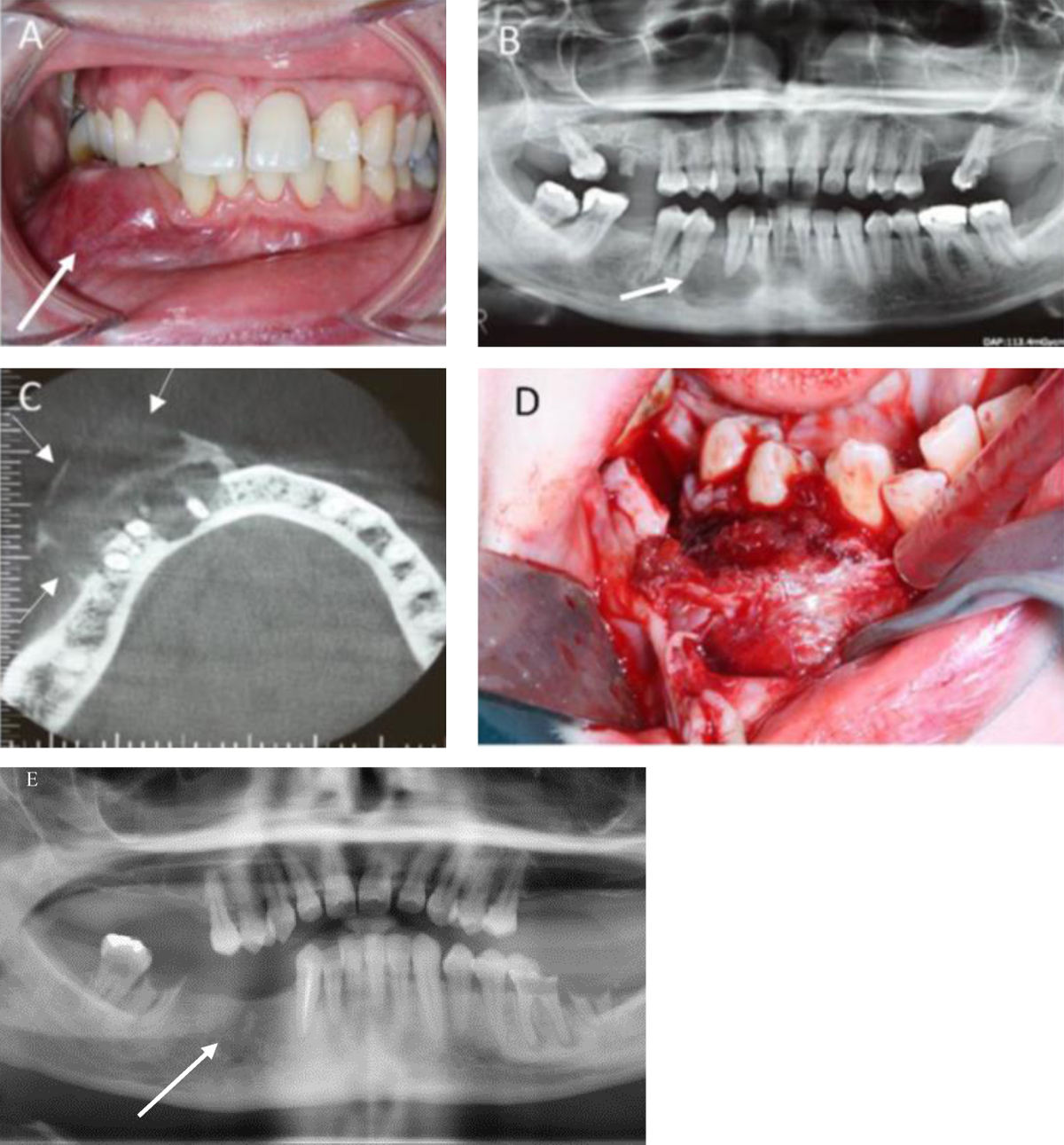
(A) Intraoral view showing a lower right vestibular swelling. (B) Orthopantogram showing the lesion at the apex of tooth 43. (C) Axial section of the cone-beam computed tomography showing the vestibular extension with a blister and perforation of the cortex and the partitions of the lesion. (D) Surgical image showing the tumorous nature of the lesion. (E) Orthopantogram of the patient 8 years after the procedure shows reossification of the site. No recurrence has occurred.
Current usage metrics show cumulative count of Article Views (full-text article views including HTML views, PDF and ePub downloads, according to the available data) and Abstracts Views on Vision4Press platform.
Data correspond to usage on the plateform after 2015. The current usage metrics is available 48-96 hours after online publication and is updated daily on week days.
Initial download of the metrics may take a while.