| Issue |
J Oral Med Oral Surg
Volume 28, Number 1, 2022
|
|
|---|---|---|
| Article Number | 7 | |
| Number of page(s) | 2 | |
| DOI | https://doi.org/10.1051/mbcb/2021033 | |
| Published online | 21 February 2022 | |
Images for Diagnosis
Breast carcinoma metastases to the jawbones: a diagnosis challenge!
1
Aix Marseille Univ, APHM, Timone Hospital, Odontology Department, Functional Unit of Oral Surgery, Marseille, France
2
Aix Marseille Univ, APHM, Timone Hospital, Odontology Department, Functional Unit of Prosthetic, Marseille, France
3
ENT and Oral Surgery Service, Edmond Garcin Hospital, Aubagne, France
* Correspondence: This email address is being protected from spambots. You need JavaScript enabled to view it.
Received:
30
August
2021
Accepted:
16
September
2021
A 44-year-old female patient was referred after the serendipitous discovery of a lytic mandibular bone lesion (Fig. 1).
She was treated in 2013 via radiotherapy, chemotherapy and surgery for a left infiltrating ductal breast carcinoma.
Clinical examination did not reveal any suspect lesion. The second left mandibular molar was mobile and vital.
A CT scan showed a unique osteolytic lesion with ill-defined margins (Fig. 2). The biopsy revealed a metastatic deposit of malignant cells matching her ductal breast carcinoma (Fig. 3). The PET scan did not show any other lesion.
Oral physicians must look for and take into account a history of breast cancer and propose a complementary exploration at the slightest clinical sign, even if it is not very evocative. Pain, swelling, and tooth mobility are frequent but not pathognomonic symptoms of jawbones metastasis. The orthopantomogram seems to be a tool of predilection in the early diagnosis of jawbones metastasis.
Conflict of interest
The authors declare that they have no conflicts of interest in relation to this article.
Informed consent
The authors declare that informed consent has been obtained.
Ethical committee approval
The authors declare that Ethical approval not required.
Source of funding
This research did not receive any specific funding.
© The authors, 2022
 This is an Open Access article distributed under the terms of the Creative Commons Attribution License (https://creativecommons.org/licenses/by/4.0), which permits unrestricted use, distribution, and reproduction in any medium, provided the original work is properly cited.
All Figures
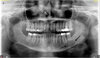 |
Fig. 1 Orthopantomogram: lytic mandibular bone lesion in the left molar area. |
| In the text | |
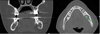 |
Fig. 2 CT scan: Unique osteolytic lesion of 19 by 11 mm of the left mandible around the first and second molar with ill-defined margins and a mandibular canal invasion. |
| In the text | |
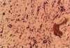 |
Fig. 3 Section from the mandibular biopsy depicting infiltrating ductal breast carcinoma. Stained with hematein, eosin, safran. Magnification ×400. 1: bone 2: malignant cells 3: non malignant cells 4: stroma. |
| In the text | |
Current usage metrics show cumulative count of Article Views (full-text article views including HTML views, PDF and ePub downloads, according to the available data) and Abstracts Views on Vision4Press platform.
Data correspond to usage on the plateform after 2015. The current usage metrics is available 48-96 hours after online publication and is updated daily on week days.
Initial download of the metrics may take a while.