| Issue |
J Oral Med Oral Surg
Volume 27, Number 1, 2021
|
|
|---|---|---|
| Article Number | 13 | |
| Number of page(s) | 5 | |
| DOI | https://doi.org/10.1051/mbcb/2020049 | |
| Published online | 07 December 2020 | |
Up-to Date Review And Case Report
Current indications of mandibular condyle plate osteosynthesis: two cases reports
1
Centre de chirurgie orale, Hôpital privé des côtes d'Armor, Plérin, France
2
Centre de chirurgie cervico-faciale et d'ORL, Hôpital privé des côtes d'Armor, Plérin, France
* Correspondence: This email address is being protected from spambots. You need JavaScript enabled to view it.
Received:
8
April
2020
Accepted:
26
August
2020
Abstract
Introduction: Certain benign or malignant pathologies may require a mandibulectomy with resection of the condyle. The gold standard for this type of reconstruction is the microanastomosed fibular free flap. An immediate reconstruction technique using an osteosynthesis plate with condyle can be proposed. The aim of this article is to evaluate the indications and describe the complications of osteosynthesis plates with condyle, through the presentation of two clinical cases. Observation: Two patients were treated by radical hemi-mandibulectomy, with cervical curage, placement of an osteosynthesis plate with condyle, and coverage with flap of pectoralis major. Then they received radiation therapy with or without chemotherapy. No cases of erosion of the glenoid fossa or tympanal bone were found. Comments: Condyle osteosynthesis plates are an interesting alternative when a fibular flap cannot be performed for local (arteriopathy), carcinologic (poor prognosis), or general and anesthesic reasons. A tissue preservation protocol, with conformation and coverage of the plate must be undertaken to limit the risk of complications (infections, exposure and fracture of the plate, temporal bone erosion, heterotopic bone formations). Conclusion: Condyle osteosynthesis plates restore aesthetic and function immediately, temporarily or even permanently. Clinical and radiological follow-up must be established following this type of reconstruction.
Key words: Osteosynthesis plate / mandibular condylar / mandibular reconstruction / case report
© The authors, 2020
 This is an Open Access article distributed under the terms of the Creative Commons Attribution License (https://creativecommons.org/licenses/by/4.0), which permits unrestricted use, distribution, and reproduction in any medium, provided the original work is properly cited.
Introduction
Many bone diseases, often fortuitously discovered, may require a radical hemimandibulectomy involving the condyle. They may be benign (such as ameloblastoma, giant cell granuloma, osteomyelitis, and ameloblastic fibroma) or malignant (such as epidermoid carcinoma, Edwing's sarcoma, and rhadomyosarcoma). In this study, we shall describe two cases involving the use of reconstruction plates and condylar prosthesis and elaborate on the elements required for the success of such a treatment.
Observations
Following extensive assessments and multidisciplinary consultations, two patients (A and B) underwent radical hemimandibulectomies with condylectomies, neck dissection, and reconstruction using condyle reconstruction plates covered with a pectoralis major flap.
Details
Patient A, a 49-year-old baker with no surgical or carcinological medical history, was suffering from type II insulin-dependent diabetes with ophthalmological and plantar comorbidities. His medical history revealed a smoking addiction (15 packs/year) as well as alcoholism (2–3 glasses daily). He initially consulted for a left laterocervical swelling that was associated with endobuccal pains. The biopsy confirmed the presence of an epidermoid carcinoma of the left mandible distal to 34 and extending up to the anterior tonsillar pillar.
Patient B, a 70-year-old retiree afflicted with hypercholesterolemia and 60% carotid stenosis without surgical indication, was being treated by his dental surgeon for a left paramandibular mass, which had appeared about 6 months prior and was sensitive to pressure from below a posterior bridge.
Diagnosis
Patient A was receiving treatment for an epidermoid carcinoma T4N2aM0 found on the floor of the mouth, which was invading the left ascending mandibular ramus. The CT scan showed that the left mandible had a lytic appearance associated with ipsilateral lymph node involvement (Fig. 1). The MRI revealed an infiltration of the masseter muscle of the furrow between the tonsil and tongue with a T1 hypointense lesion on the left mandible. No perineural infection of the third branch of the trigeminal nerve was present at the level of the foramen ovale.
Patient B presented with a cystic, mandibular mass of pseudo-fistular appearance with bony arches. The CT scan revealed a bilobed lesion to the posterior part of the ascending ramus with bilateral bony fenestrations. The diagnosis of an ameloblastic carcinoma, class T4N0M0, was only possible after the histological analysis of the excised tissue.
 |
Fig. 1 Preoperative CT scan: 3D reconstruction (Patient A). |
Intervention
During a multidisciplinary consultation meeting, a surgical strategy was proposed for patient A, comprising an interruptive hemimandibulectomy, including the condyle, cervical and ipsilateral dissection, and reconstruction by reconstruction plates covered with a pectoralis major flap (Fig. 2). A free flap was not feasible due to the patient's smoking history, insulin-dependent diabetes complicated by retinopathies, the existence of a neuropathic ulcer, and peripheral arteriopathies that were confirmed by echo-Doppler scanning. Following the intervention, a dosage of 66 Grays (Gy) were administered to the tumor bed and lymph nodes by tumor conformational irradiation at modulated intensity; this was combined with adjuvant chemotherapy comprising three cycles of cisplatin 100 mg/m2 every 3 weeks.
For patient B, an excision with preservation of the bone and inferior alveolar nerve was first conducted under general anesthesia. A fibrous mass associated with a hematoma had caused shrinkage of the fenestrations of the inner cortex of the mandible (Fig. 3). The histological analysis of the excised tissue also revealed the malignant character of the tumor; a radical surgical treatment by interruptive mandibulectomy, cutting across healthy bone, was performed in the following weeks.
The plate was shaped with a template of the same height and length as the excised tissue. An intermaxillary locking mechanism was necessary to attach the plate with the screws extending into the contralateral chin area to establish the patient's preoperative occlusion (Fig. 4). The temporomandibular joint capsule and the articular disc were preserved, and the plate was suspended in the joint with non-absorbable wires. The plate was covered with a pectoralis major flap. To avoid postoperative discharge tracheostomy, the patient remained intubated for 24 h after the intervention. The intermaxillary locking mechanism was prolonged for 15 days. An irradiation dose of 70 Gy was administered without combined chemotherapy.
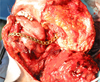 |
Fig. 2 Mandibular reconstruction plate with prosthetic condyle and pectoralis major flap (Patient A). |
 |
Fig. 3 Ameloblastic carcinoma (Patient B): preoperative orthopantomogram (left) and excision tissue (right). |
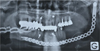 |
Fig. 4 Postoperative orthopantomogram with reconstruction plate and intermaxillary locking mechanism (Patient B). |
Therapeutic follow-up
After 2 years of postoperative monitoring, patient A died of related cardiopulmonary and diabetic complications. During his last consultation it was noted that the plate was well-tolerated and that the cancer was in remission.
Patient B's 5-year scan showed that the plate was perfectly integrated into the glenoid fossa without any sign of temporal bone erosion (Fig. 5). Moreover, no infectious complications or exposure of the osteosynthesis materials were observed. The clinical and radiographic follow-up of the patient continued for 12 years, and he has retained satisfactory levels of masticatory function and phonation as well as an aesthetic facial appearance (Fig. 6).
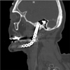 |
Fig. 5 Five-year follow-up CT showing the integrity of the glenoid fossa against prosthetic condyle (Patient B). |
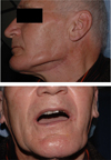 |
Fig. 6 Five years follow-up photographs (Patient B). |
Discussion
Indications of plate osteosynthesis with condylar prosthesis
While performing a hemimandibulectomy, condylar reconstruction remains a major surgical challenge. After the excision of the mandibular tumor with condylectomy, several secondary reconstructive techniques have been documented with or without prosthetic materials. These reconstructions are performed with materials such as full titanium condylar plates [1,2], non-vascularized bone flaps with costochondral sampling [3], iliac, or a micro-anastomosed, free, fibular flap [4]. The progress made in reconstructive surgery, especially concerning vascular anastomoses in microsurgery, constitutes the gold standard of condylar reconstructions. This type of flap presents certain advantages, particularly its stability and resistance to infections, and enables the placement of implants on fibular bone grafts.
However, attaching the plate to the condyle is also indicated in certain situations. Thus, it is an interesting alternative during peripheral arteriopathies (patient A) where prolonged general anesthesia is not supported and where the carcinological prognosis is unfavorable [4]. Titanium prostheses aesthetically as well as functionally facilitate the proposal of immediate mandibular reconstruction. They require a lighter technical platform and a single operating team, unlike the anastomosed flaps. They are nonetheless contraindicated in cases where the lesion invades the mandibular symphysis and in children. Some teams have noted the lack of reconstructions in cases of consecutive mandibular deviation on the operated side [5].
Shaping and positioning of the plate
Several systems for condylar plate osteosynthesis exist and have been proposed by many manufacturers, including Synthes™ (Paoli, USA), Stryker Leibinger™ (Kalamazoo, USA), and KLS Martin™ (Tuttlingen, Germany).
The plate must conform to the initial length of the ramus as well as the mandibular angle; therefore, a shaped malleable template is used and measured against the excised tissue to reestablish facial symmetry and to harmoniously redistribute the occlusal forces. The condylar head must be ligatured (non-absorbable synthetic wire) and suspended amidst the adjacent muscles to prevent its movement and improve its stability. Care must also be taken to reform the masseteropterygoid belt by suturing the muscles along the angle of the plate. The proper shape of the plate also prevents any pressure on the temporal bone during occlusion that may cause bone resorption [6]. An inter-maxillary locking mechanism is recommended because it stabilizes the dental articulator while the wound is healing.
The need to interpose a material between the plate and the temporal bone
Of all the complications associated with prosthetic condylar plate reconstruction, temporal resorption and movement toward base of the skull remain the most serious. The excision of the temporomandibular joint capsule and articular disc constitutes an absolute contraindication to metallic restoration alone [6]. Effectively, direct contact between the temporal bone and the metallic condylar may lead to bone resorption of the glenoid cavity and movement of the plate subsequent to which the overlying structures will be affected. Described among the latter are tympanic bone lesions and cranial fossae [7]. Cochlear lysis causing neurosensory deficits is also described [8]. These resorptions are sometimes accompanied by heterotopic bone formations around the condylar head [9]. The interposition of adipose tissue, cartilage, or muscular fascia no longer constitutes a viable solution because secondary dehiscence can lead to contact with the glenoid cavity. Furthermore, Desai and Smit propose the use of a temporal plate made of high-density polyethylene (Zimmer Biomet CMF™, Jacksonville, Florida, USA) as a means of supporting the metallic condylar prostheses. They applied this protocol 12 weeks after surgery among nine patients who presented with no clinical and radiological pathological signs [10].
Covering the plate with soft tissue
Although certain advantages exist for using the plates during mandibulectomies with condylar excision, these procedures are still not exempt from complications. Secondary radiotherapy can cause infections with cutaneous and mucosal fistulation that could result in its externalization [11,12]. Therefore, it is advised that the plate is covered with soft tissue to limit the risk of exposure, restore volume, and make aesthetic improvements. It can be covered with a muscular pedicle flap (temporal or sternocleidomastoid) or musculocutaneous (pectoralis major or dorsalis major) or even free (major muscle straight from abdomen) [6]. The flap of the pectoralis major is indicated for this type of resection since it is musculocutaneous, pedicled, and vascularized with only one operation site and since it does not require microanastomsosis. Moreover, it has an established clinical track record [2].
Success rate and long-term follow-up (Tab. I)
Some studies favor reconstruction platesthat, according to the authors, are only transitional techniques before the bone graft [8,13]. However, the authors did not cover the plate with a flap and had interposed the prosthetic condylar in direct contact with the temporal bone. Other studies have found a higher success rate [1,2,6,10,11]. In one retrospective study in 2003 that examined six cases, of which four involved chemotherapy and an average follow-up of more than 2 years, there were no failed cases when the metallic condylar prostheses was used [6]. A 2008 prospective study (Marx et al.) examining 131 cases with a follow-up period of more than 2 years reported a complication rate of 10.6% with only one reported case of erosion of the external auditory canal, two cases of plate loss, and six cases of exposure, exclusively in irradiated patients who had no covering flap. They concluded that the plate does not necessarily have to be deposited if it is stable and functional [11]. By comparison, the average complication rate at 3 years with a fibular flap is 20.2% [14]. Huang et al. in a 2015 study examining 12 cases reported a complication rate of 41.7% within the 3 to 5 years post-reconstruction, of which one case involved the erosion of the glenoid fossa, two cases reported the displacement and erosion of the external auditory canal, and other complications that are not detrimental to the survival of the material [15].
Literature review of reconstruction plates with condyle.
Therapeutic perspectives
Reconstruction plates with condylar prosthesis are an interesting choice for immediate restorations because they limit morbidity, reduce post-operational side effects, and restore mandibular function. The plate must be placed as close as possible to the initial mandibular anatomy and must be covered by a local flap to limit the risk of exposure and restore volume to the resected area. The condylar head must be stabilized without coming into direct contact with the temporal bone. At present, 3D modeling techniques can help with reconstruction; however, the final adjustment of the plate falls under the technical expertise of the surgeon.
Conflicts of interests
None.
Acknowledgements
The authors would like to thank the imaging and oncology team at the private hospital in Cotes d'Armor, Pontchaillou University Hospital Centre, Rennes. We would also like to thank the Eugène Marquis Center for their help in preparing this study.
References
- Vuillemin T, Raveh J, Sutter F. Mandibular reconstruction with the titanium hollow screw reconstruction plate (THORP) system: evaluation of 62 cases. Plast Reconstr Surg 1988;82:804–814. [PubMed] [Google Scholar]
- Vuillemin T, Raveh J, Sutter F. Mandibular reconstruction with the THORP condylar prosthesis after hemimandibulectomy. J Cranio-Maxillo-fac Surg Off Publ Eur Assoc Cranio-Maxillo-fac Surg 1989;17:78–87. [Google Scholar]
- Lindqvist C, Jokinen J, Paukku P, Tasanen A. Adaptation of autogenous costochondral grafts used for temporomandibular joint reconstruction: a long-term clinical and radiologic follow-up. J Oral Maxillofac Surg 1988;46:465–470. [PubMed] [Google Scholar]
- Lin C-H, Kang C-J, Tsao C-K, Wallace CG, Lee L-Y, Lin C-Y, C et al. Priority of Fibular reconstruction in patients with oral cavity cancer undergoing segmental m and ibulectomy. Elsalanty M, Ed. PLoS ONE 2014;9:e94315. [Google Scholar]
- Mallet Y, Lefebvre JL. Oropharyngeal surgery (bucco-pharyngectomies). Medico-surgical encyclopedia. Surgical Techniques − Head and Neck 2008;1-10:46-320. [Google Scholar]
- Daniel E. Minimizing complications in the use of titanium condylar head reconstruction prostheses. Otolaryngol Head Neck Surg 2004;130:344–350. [PubMed] [Google Scholar]
- Carlson ER. Disarticulation resections of the mandible: a prospective review of 16 cases. J Oral Maxillofac Surg 2002;60:176–181. [PubMed] [Google Scholar]
- Patel A, Maisel R. Condylar prostheses in head and neck cancer reconstruction. Arch Otolaryngol Head Neck Surg 2001;127: 842-846. [PubMed] [Google Scholar]
- Lindqvist C, Söderholm AL, Hallikainen D, Sjövall L. Erosion and heterotopic bone formation after alloplastic temporomandibular joint reconstruction. J Oral Maxillofac Surg Off J Am Assoc Oral Maxillofac Surg 1992;50:942–949. [Google Scholar]
- Desai J. The use of reconstruction plates and add-on condyles with an alloplastic unmatched fossa, following partial mandibulectomy with disarticulation. J Surg 2019;7:31-34. [Google Scholar]
- Marx RE, Cillo JE, Broumand V, Ulloa JJ. Outcome analysis of mandibular condylar replacements in tumor and trauma reconstruction: a prospective analysis of 131 cases with long-term follow-up. J Oral Maxillofac Surg 2008;66:2515–2523. [CrossRef] [PubMed] [Google Scholar]
- Kim MR, Donoff RB. Critical analysis of mandibular reconstruction using AO reconstruction plates. J Oral Maxillofac Surg Off J Am Assoc Oral Maxillofac Surg 1992;50:1152–1157. [Google Scholar]
- Westermark A, Koppel D, Leiggener C. Condylar replacement alone is not sufficient for prosthetic reconstruction of the temporomandibular joint. Int J Oral Maxillofac Surg 2006;35:488–492. [PubMed] [Google Scholar]
- Wu H, Liu F, Ji F, Guo M, Wang Y, Cao M. Identification of independent risk factors for complications: a retrospective analysis of 163 fibular free flaps for mandibulofacial reconstruction. J Oral Maxillofac Surg 2018;76:1571–1577. [PubMed] [Google Scholar]
- Huang J-S, Chen W-H, Liu P-H, Huang T-T, Hsiao J-R, Ho C-L. Outcome analysis of condylar prothesis in large head and neck neoplasm reconstruction. J Mech Med Biol 2015;15:1–10 (1540040). [Google Scholar]
All Tables
All Figures
|
Fig. 1 Preoperative CT scan: 3D reconstruction (Patient A). |
| In the text | |
|
Fig. 2 Mandibular reconstruction plate with prosthetic condyle and pectoralis major flap (Patient A). |
| In the text | |
|
Fig. 3 Ameloblastic carcinoma (Patient B): preoperative orthopantomogram (left) and excision tissue (right). |
| In the text | |
|
Fig. 4 Postoperative orthopantomogram with reconstruction plate and intermaxillary locking mechanism (Patient B). |
| In the text | |
|
Fig. 5 Five-year follow-up CT showing the integrity of the glenoid fossa against prosthetic condyle (Patient B). |
| In the text | |
|
Fig. 6 Five years follow-up photographs (Patient B). |
| In the text | |
Current usage metrics show cumulative count of Article Views (full-text article views including HTML views, PDF and ePub downloads, according to the available data) and Abstracts Views on Vision4Press platform.
Data correspond to usage on the plateform after 2015. The current usage metrics is available 48-96 hours after online publication and is updated daily on week days.
Initial download of the metrics may take a while.