Fig. 1
Download original image
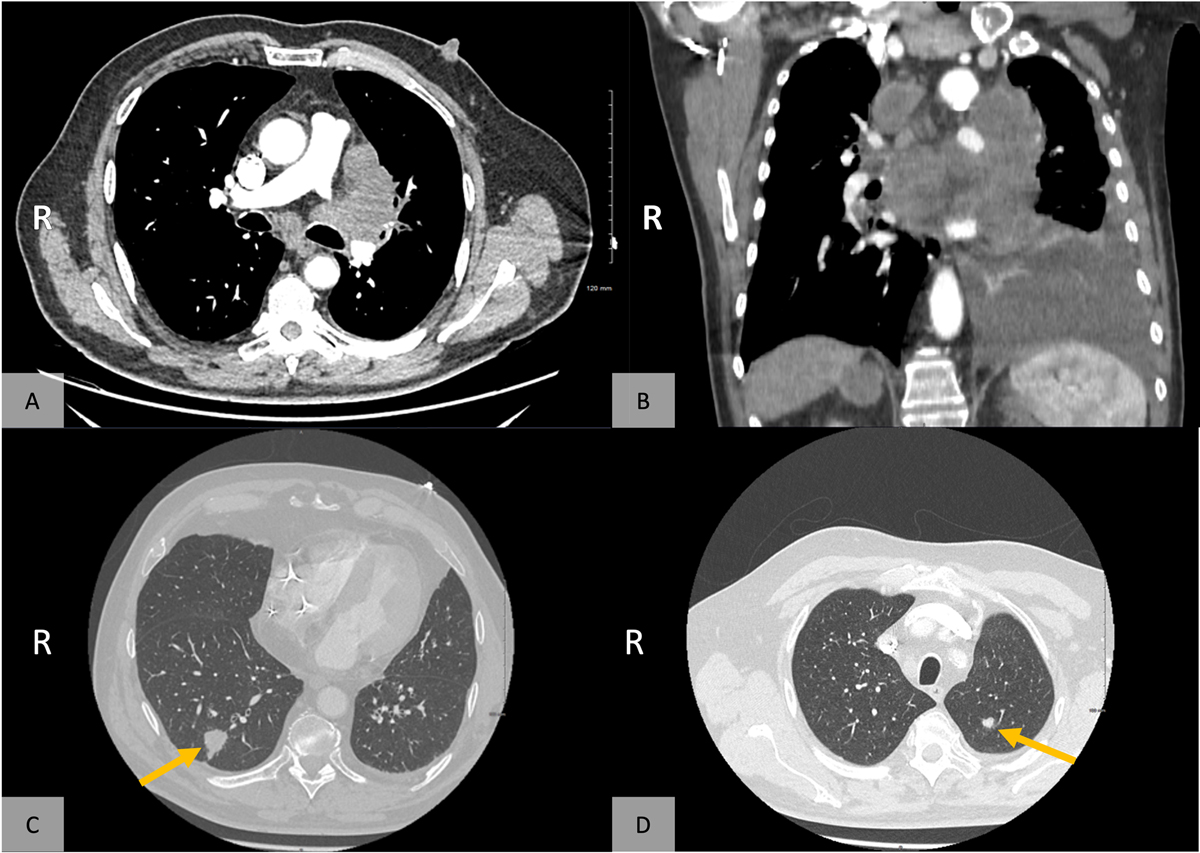
(A) Axial mediastinal window CT sections show large mediastino-hilar masses, more voluminous at the left and in subcarinal area, left pleural effusion. (B) Coronal mediastinal window CT section. (C-D) Axial lung window CT sections show a postero-basal spiculated nodule of secondary appearance in right lower pulmonary lobe (C), and another one in left apical upper lobe (D) (yellow arrows).
Current usage metrics show cumulative count of Article Views (full-text article views including HTML views, PDF and ePub downloads, according to the available data) and Abstracts Views on Vision4Press platform.
Data correspond to usage on the plateform after 2015. The current usage metrics is available 48-96 hours after online publication and is updated daily on week days.
Initial download of the metrics may take a while.