Fig. 4
Download original image
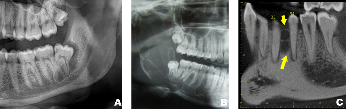
Radiographic features of keratinized odontogenic cysts. (A) Panoramic view showing an unilocular radiolucency surrounding the distal aspect of a partially erupted molar. The final diagnosis was an orthokeratinized odontogenic cyst. (B) Panoramic view showing an extensive multilocular radiolucency involving the right ascending ramus. The final diagnosis was an odontogenic keratocyst. (C) Sagittal section of a CBCT showing a multilocular radiolucency in a “lateral periodontal” emplacement. The final diagnosis was surprisingly a keratocyst.
Current usage metrics show cumulative count of Article Views (full-text article views including HTML views, PDF and ePub downloads, according to the available data) and Abstracts Views on Vision4Press platform.
Data correspond to usage on the plateform after 2015. The current usage metrics is available 48-96 hours after online publication and is updated daily on week days.
Initial download of the metrics may take a while.