Fig. 11

Download original image
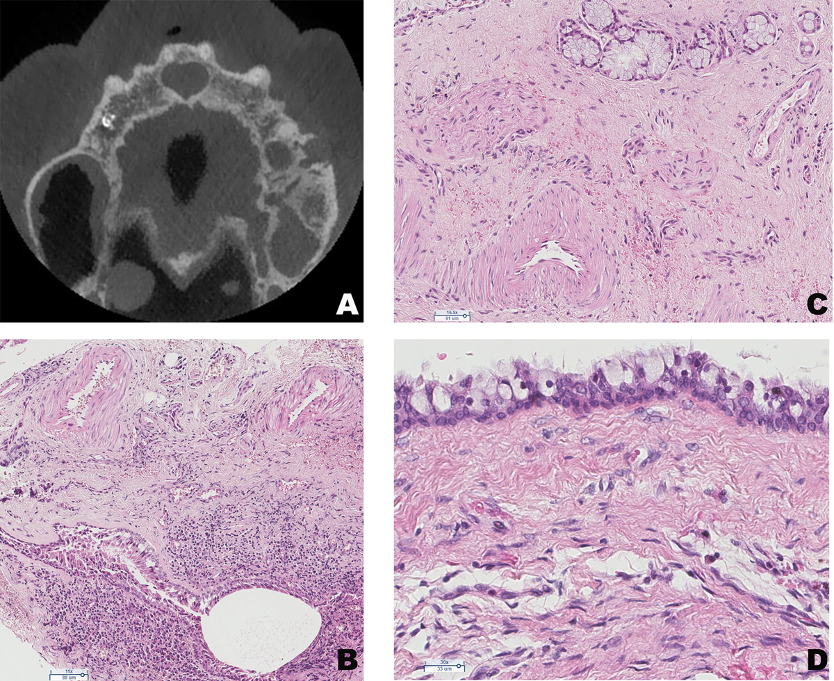
Radiological and histological features of a nasopalatine duct cyst. (A) CBCT horizontal section showing a “heart-shaped” radiolucency between superior incisors. (B) Pseudostratified cystic epithelium and prominent arteries within the cystic wall (HE ×110). (C) Mucous gland and prominent neurovascular bundles within the cystic wall (HE ×165). (D) Presence of mucous cells within the epithelium of a radicular cyst : these cells are not specific of any odontogenic cysts (HE ×300).
Current usage metrics show cumulative count of Article Views (full-text article views including HTML views, PDF and ePub downloads, according to the available data) and Abstracts Views on Vision4Press platform.
Data correspond to usage on the plateform after 2015. The current usage metrics is available 48-96 hours after online publication and is updated daily on week days.
Initial download of the metrics may take a while.


